When Screens Replace Connection: Impact on Children’s Brains
Posted: August 31, 2026 Filed under: ADHD, attention, behavior, cellphone, computer, emotions, Evolutionary perspective, Exercise/movement, healing, health, laptops, lifestyle, neurofeedback, screen fatigue, techstress, Uncategorized | Tags: infant development, neurodevelopment, screeen trauma, screen addiction, serve and return, snesory deprivation, social engagement, synesthesia, touch 1 CommentErik Peper, PhD, BCBa, Stoyan Vezenkov, PhDb, Violeta Manolovab, Lauren Mason, PhDc and Richard Harvey, PhDd
a BiofeedbackHealth, Berkeley, CA; b Center for Applied Neuroscience Vezenkov, Sofia, Bulgaria; c Mason-White Consulting, La Mesa, CA, USA; d San Francisco State University, San Francisco, CA, USA

Why are top tech CEOs strictly limiting their own children’s screen time while digital devices increasingly dominate classrooms and homes? In this latest post, When Screens Replace Connection: The Impact on Children’s Brains, we examine the physiological realities behind the growing mental-health challenges facing young people. We explore how excessive screen exposure may contribute to traits associated with ASD, ADHD, and emotional dysregulation, including through mechanisms such as “screen-induced dysregulation” and the erosion of vital “serve-and-return” interactions between children and caregivers.
Drawing on psychophysiological research, the article investigates how digital media can disrupt the human “social engagement system.” When face-to-face eye contact, vocal prosody, physical touch, movement, and responsive interaction are replaced by rapid 2D stimulation, developing systems involved in attention, attachment, and stress regulation may be affected.
It includes practical, evidence-based strategies for families, educators, and communities to restore balance through embodied connection, shared presence, movement, and time in nature. Rather than rejecting technology altogether, we advocate for more intentional use: fewer dependency-forming screens and more opportunities for responsive caregiving and real-world connection. The blog asks an essential question: Are we giving children more digital stimulation than human connection—and what might they be losing as a result?
*********************************
“So, your kids must love the iPad?” I asked Mr. Jobs, trying to change the subject. The company’s first tablet was just hitting the shelves. “They haven’t used it,” he told me. “We limit how much technology our kids use at home” (Bilton, 2014).
“Peter Thiel and other tech billionaires are publicly shielding their children from the products that made them rich” (Quiroz-Gutierrez, 2026).
Many of us are aware that something has shifted in the way we live, learn, and relate to one another. Screens—once tools of convenience—have become our primary portals for information, education, and social connection. Yet this shift comes with an unintended cost. The social psychologist Jonathan Haidt (2024) compellingly documents in his book, The Anxious Generation, that the rapid rise in screen use, especially social media content, parallels a striking decline in the self-reported mental health of young people (Haidt, 2024).
Beginning in the early 2000s, general rates of anxiety, depression, loneliness, and self-harm among adolescents have increased (Haidt, 2022). The trend did not start with the COVID-19 pandemic; rather, it preceded it by more than a decade. Facebook was launched in 2004, and the iPhone—ushering in the era of constant connectivity—arrived in 2007. Social media content combined with omnipresent technological innovations fundamentally reshaped how people, and especially young people experience the world: less face-to-face interaction, increasingly curated online identities, as well as a constant stream of social comparisons and forms of stimulation aimed at engaging developing brains (Swingle, 2019).
From a psychophysiological perspective, human beings evolved to regulate physical stress along with their emotional reactions without social media content. Instead the human ‘social engagement system’ linking heart, lungs and facial expressions (e.g. Ventral Vagal Complex) (Porges, 2011) along with direct social contact that includes eye gaze, touch, and shared presence regulate stress and emotional reactions such as release of hormones (e.g. vasopressin and oxytocin) (Eckstein, Mamaev, Ditzen, & Sailer, 2020; Atzil et al., 2018). When ‘social engagement’ behaviors are replaced by screen-mediated interactions, the body loses key cues for safety and connection (Haidt, 2022). The result can be a subtle but chronic activation of stress responses, contributing to emotional dysregulation, sleep disruption, reduced resilience, and an increase in serious psychologic distress, depression and suicidal thoughts, as shown in Figure 1 (Braghieri et al, 2022).
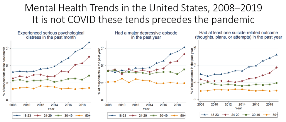
Figure 1. Mental health trends in the United States by age group in 2008–2019. The data come from the National Survey on Drug Use and Health. From: Braghieri, L., Levy, R., & Makarin, A. (2022). Social media and mental health. American Economic Review, 112(11), 3660–3693. https://doi.org/10.1257/aer.20211218
As screens become more prevalent and are increasingly incorporated into TK–12 education, with some municipalities enrolling children as young as 3.5 years of age, the promise of improved social engagement learning often fails to be realized. There is a parallel in reduced levels of intellectual attainment. For most students—outside of small groups who already perform at very high levels—math and reading scores have steadily declined since the introduction of social media content post COVID-2019 (Kus, 2025). In the United States, math scores are at their lowest levels in decades, according to the National Assessment of Educational Progress (NAEP). There was also a meaningful decline in overall science scores for 8th graders, with almost 40% of students below a ‘basic’ level of proficiency and a similar decline in math and reading scores for 12th graders in 2024 compared to 2019 representing what has been called the largest achievement gap in decades (NAEP, 2022; Harris, 2025),.
The findings related to deficits of social engagement and academic achievement do not mean that technology use is inherently harmful. Rather, they highlight a mismatch between our biological needs and our digital habits of engaging with dependency-forming content delivered by technology. Just as we learned to balance nutrition, movement, and rest for our well-being, we now face the challenge of consciously shaping our relationship with screens (Peper et al., 2020). Reintroducing embodied experiences— movement and dance, real-world social interactions, and time spent with others in nature—can help restore physiological balance and emotional flourishing by amplifying the social engagement system which evolved over millennia.
Although some have suggested that screen addiction may be overestimated (Anderson & Wood, 2025), many people report concern about the effects on young adults from exposure to digital dependency-forming content, this content delivered on screens and rapid digital displays may be even more harmful to infants and toddlers (Amirthalingam, & Khera, 2024; Reinecke, Gilbert, & Eden, 2022). The brains of young children are still developing and forming new neural connections and pathways in response to these ‘digital-engagement’ stimuli. There are two major risk factors:
- Caregivers being physically present but not attending to or communicating with the infant or toddler, and
- The remodeling of infants’ and toddlers’ brains through exposure to screens and their content.
- Caregivers being physically present but not attending to or communicating with the infant or toddler
Being physically nearby a child however attending to content on a screen rather than attending to the infant or toddler results in potential for experiencing feeling left out, ignored or abandoned and misses opportunities for personal, dynamic, interactive social feedback. Analogously, there may be a process that is similar for college students who report feeling neglected when they are engaged in a conversation that becomes interrupted by a phone notification (Peper & Harvey, 2018).
Infant development and communication are a dyadic feedback process (e.g., behavioral and neural synchronization or coupling; the “Serve and Return” Framework) which is essential for healthy brain architecture in which the caregiver and infant respond to each other rather than to digital content (Carozza & Leong, 2021; Ilyka, Johnson & Lloyd-Fox, 2021; Theyer & Wijeakumar, 2025). Because brains continue to mature for at least two or more decades, college students are likely also benefitting from increased social engagement rather than digital media engagement.
For example, dynamic social engagement is often studied through the lens of serve-and-return interactions (Chen et al., 2023; Komanchuk et al., 2023). If communication pathways are not activated, then the biological foundation for social and emotional skills may not develop fully (Shonkoff & Phillips, 2000). Infants and young children may learn that their needs are not consistently met, leading to “avoidant” or “disorganized” attachment styles. This can manifest later in life as difficulty trusting others or difficulty regulating one’s own emotions (Gunnar & Quevedo, 2007). Spoken languages and social cognitions are learned through mirroring and feedback loops. Without active participation during language acquisition—when babies learn the rhythm of conversation and the meaning of sounds by watching their caregivers’ responses—their vocabulary growth and verbal communication are often significantly delayed. These children may also lack social understanding as they can struggle to interpret social cues or develop empathy later on (Kuhl, 2007; National Scientific Council on the Developing Child, 2012; National Scientific Council on the Developing Child, 2026).
When a caregiver does not respond to the subtleties of the infant’s communication, the infant may be unable to learn effectively and may also feel frustrated. The developmental harm of inattention while physically present has been graphically shown by the research of Dr. Edward Tronick in the Still Face Experiment, as demonstrated in the following two videos (Still face with mother: https://www.youtube.com/watch?v=YTTSXc6sARg ; Still face with dad: https://www.youtube.com/watch?v=7Pcr1Rmr1rM(Tronick, 2007; Weinberg, et al., 2008).
The extreme harm of neglect along with lack of social engagement including non-verbal feedback has previously been demonstrated in the research by Harry Harlow’s 1950s–1960s experiments on rhesus monkeys, who were placed with wire or cloth surrogate mothers rather than real mothers. These studies showed that infant attachment is initially and primarily driven by comfort and warmth interactions. More importantly, adult monkeys, when raised in isolation or with artificial mothers, were socially incompetent, often unable to interact or mate, and exhibited severe behavioral issues (Harlow et al., 1965).
Similar findings have been observed with human infants. For example, following the political transfer and fall of Romanian leader Nicolae Ceaușescu in 1989, it was revealed that over 100,000 children in Romanian institutions faced extreme physical, cognitive, and emotional neglect. Confined to cribs with minimal human contact, these babies suffered severe developmental delays, structural brain changes, and profound attachment issues, often exhibiting self-stimulatory behaviors (Nelson et al., 2023).
It is important to acknowledge that parents and caregivers do not typically go to the extremes seen in Harlow’s rhesus monkeys or the neglected Romanian infants; however, implicit neglect can occur when the caregiver is not attentive to their children and are instead absorbed in their screen content. Engaging in dynamic serve-and-return interactions with the infant reduces a risk factor for healthy infant development. The solution is obvious: attend to and interact with people, especially infants and young children in a serve-and-return style. When interacting with infants, be present:
- Put your cellphone away and engage dynamically with the infant and children.
- Do not give a screen to an infant as a distraction. Instead, play with and attend to the child while sharing responsibilities as primary caregivers with others in a household or community.
2. The remodeling of infants’ and toddlers’ brains through exposure to screens
Over the past decade, through their clinical and research work at the Vezenkov Center for Applied Neuroscience in Sofia and Weinheim, Drs. Vezenkov and Manolova have arrived at a position that may sound strong at first hearing but which, in our view, is now unavoidable: in a substantial subgroup of young children presenting with severe disorders with features that are like autism spectrum disorder (ASD-like), Attention Deficit Hyper-Activity Disorder (ADHD-like), Oppositional Defiant Disorder (ODD-like), or Pervasive Developmental Disorder (PDD-like) features, there are no observations of a primary neurodevelopmental disorder. Rather there is a clinical signature of early screen addiction (Vezenkov & Manolova, 2025a), screen trauma (Manolova & Vezenkov, 2025a), and may be called ‘reversed development’ (Manolova & Vezenkov, 2025c). Importantly, treating these children for screen addiction and screen trauma, the clinical picture changes, and in many cases the original ASD or ADHD diagnosis is lifted. This is a clinical hypothesis derived from repeated observations and consistent therapeutic outcomes — offered as a framework that is testable, falsifiable, and one that can be described as clinically urgent.
Screen addiction is not screen time
The first conceptual step is to separate screen addiction from screen time. Severe early screen addiction is not simply indexed by hours in front of a device. Rather, (a) compulsive seeking of high-intensity, low-social-density visual or sensory stimulation combined with (b) cycles of soothing and crash as well as (c) a shift of interest away from human faces toward objects, colors, shapes, and repetitive sensory patterns along with (d) a collapse of joint attention and eye-to-eye play. Removing the device produces withdrawal — crying, hysteria, aggression, or shutdown — not because the child is willful, but because what has been removed is the child’s regulatory prosthesis.
Three learning systems (e.g. identified as ‘System 0, 1 and 2 described below) frame and organize behavior during clinical care. Drawing on Kahneman, System 2 is the slow human system: language, reflective thinking, social context, values, and the capacity to delay action (Kahneman, 2011; Khalil & Brüne, 2025). System 1 is the fast system: automatisms, immediate reward, and reinforcement through the dopaminergic reward circuitry. A third is System 0, drawing on Rizzolatti’s work on mirror neurons — the biological bridge between “I see” and “I do” in human encounters (Rizzolatti & Sinigaglia, 2016). In healthy development, the three systems work together. In screen-dominant development, System 1 wins and closes an addiction loop, System 2 stalls, and System 0 is hacked: human mirroring is replaced by immersion in the object world. Mirror neurons are not damaged in these children; they are retuned, away from faces and toward screens.
The developing brain: sensitive windows and the arrest of pruning
The first six years of life are a period of intense synaptogenesis, reorganization, and myelination. The brain calibrates itself to the environment — deciding which sounds are meaningful, which faces matter, and which rhythms feel safe. The dominant cortical frequency accelerates from delta in the newborn to roughly 10 Hz at age nine, at approximately one hertz per year (Freschi et al., 2022; Wilkinson et al., 2024). Behind this gradient lies a series of sensitive windows in which the child’s experiences shape what the brain becomes.
When the environment is rich in human movement, touch, eye contact, prosody, and rhythm, the brain prunes excess connections and strengthens those that support attention, language, and self-regulation. When the environment is dominated by fast, repetitive audiovisual stimulation, the brain strengthens those pathways instead. The auditory and visual sensory systems become hypertrophic at the expense of executive motor loops, affect, and speech, producing the splitting and fragmentation of cortical function described below.
A second key point is pruning. The hypermnesic period around 18–36 months should normally be followed by reorganization and selection (Gonzalez-Escamilla et al., 2018; Sakai, 2020). In severe early screen addiction, this process does not unfold normally. Development does not simply slow; it freezes. Children enter what we term addictive homeostasis — a state actively defended against change. In clinical settings, we observe twelve-year-olds who have not yet shed their primary teeth, as well as delayed acquisition of bowel and bladder control. The child may accumulate fragments — words, scripts, colors, routines — without integrating them into a language system or socially meaningful behavior. The result is not delay but a traumatization and deformation of development that cannot self-correct without specialized intervention.
Biomarkers: the physiological signature of screen addiction
If this framework is to be taken seriously, it must leave physiological traces. In our work it does so consistently across several hundred qEEG and HRV recordings in children, adolescents, and adults (Vezenkov & Manolova, 2025a). We observe four broad classes of biomarkers in early screen addiction:
- Altered cortical activity in opposing directions across networks. Some leads show slowing — theta and/or alpha peaks in central, frontal, and frontopolar sites — while others show acceleration, with SMR, beta1, or beta2 bursts in central, parietal, and occipital sites. Similar findings have also been reported by Law et al. (2023). The two patterns coexist and produce what we call cortical splitting. Other researchers have observed similar effects in which EEG results suggested a higher connectivity in theta vs. beta bands in the screen group, but not in the control group (Zivan et al., 2019). Their results support the negative relationship between screen exposure and attention-related patterns generated from EEG in typically developing preschool children.
- Reversed hemispheric asymmetry, with dominant alpha or theta rhythms in the left hemisphere — the inverse of the typical pattern.
- Functional fragmentation: each cortical site behaves as if running on its own clock, with frequency differences between leads often greater than 1.5 Hz. High local coherence coexists with weak long-range coherence, indicating disrupted connectivity. This pattern is most pronounced when onset of screen exposure is between 0 and 3 years.
- Autonomic dysregulation, expressed in HRV as sympathicotonia, dorsal vagotonia, or unstable mixed patterns — rather than the ventral vagal balance associated with secure attachment and social engagement (Porges, 2011; Pankova et al., 2021). In the clinic it has been observed that the age of onset of severe screen exposure leaves a signature in the dominant cortical frequencies. Onset before 18 months tends to be associated with peaks at 2–3 Hz; 18–30 months with 3–4 Hz; 30–36 months with 4–5 Hz. This remains a practice-derived hypothesis rather than an established rule, but it aligns strikingly with parental histories. The biomarker profile does not, on its own, constitute a diagnosis. Together, however, these markers outline a recognizable physiological spectrum from nonverbal dorsal-freeze states to verbal, high-functioning children with autistic traits.
Screen trauma: why detox alone is not enough
Parents and clinicians often assume that simply removing screens will return the child to a typical developmental trajectory. In many cases it does not. Even after weeks or months of complete digital detox, the child remains anchored in infantile, sensory-dominant patterns. During clinical work, we have come to call this state screen trauma (Manolova & Vezenkov, 2025a).
Screen trauma is what state-dependent learning looks like when the state in which learning was acquired was screen-induced dysregulation. The anchors are physiological: persistent primitive reflexes, atypical vestibular and postural patterns, sensory disintegration, cortical fragmentation, and a vision-dominant mode of processing. Functioning is tuned to stereotyped, repetitive stimuli — which the world outside the screen does not provide. Removing the screen removes the trigger but leaves the architecture in place. Recovery requires therapeutic work that lifts these anchors sequentially.
Pathological screen-induced reflexes: SIPVR and SIPECR
Two clinical reflexes have been identified that serve as among the most useful sensitive markers for screen trauma. Both are present in screen-addicted children and absent in typically developing peers, and both resolve with therapy and re-emerge with screen re-exposure, confirming their conditioned, environmental nature:
The Screen-Induced Pathological Vestibular Reflex (SIPVR) is elicited by a controlled backward tilt or inversion of the child (Vezenkov & Manolova, 2025c). An affected child shows arching backward, reaching out to grasp support, trembling and panic, full-body muscular stupor while desperately seeking support, and intense fear of falling — disproportionate to the postural challenge. In typically developing children, the same maneuver elicits play, laughter, or rapid adaptation. The reflex is highly sensitive in children up to age twelve. The SIPVR can be seen in the following video: https://youtu.be/_9PKtanDClU?si=KNicSRlDqEAutBQihttps://youtu.be/_9PKtanDClU?si=KNicSRlDqEAutBQ
The Screen-Induced Pathological Eye-Covering Reflex (SIPECR) is elicited by briefly covering the child’s eyes with hands, a mask, or a cloth (Vezenkov & Manolova, 2025d). Affected children respond with intense panic, screaming, agitation, and in severe cases aggression toward caregivers or self-injurious behavior such as head-banging. Episodes can persist for ten to thirty minutes and do not de-escalate while the visual occlusion remains. The SIPECR reflex was observed in 275 of 285 children evaluated at our center (96.5%). Unlike a startle reflex, it is provoked by the removal rather than the onset of stimulation; unlike nyctophobia, it persists when the child is enclosed under a translucent cloth in the presence of a caregiver, demonstrating that the trigger is not darkness itself but the loss of familiar visual cues. SIPECR is not observed in typical development and, alongside SIPVR, helps differentiate screen trauma from primary neurodevelopmental conditions.
Sensory disintegration versus sensory deprivation
These reflexes acquire their full meaning within a broader framework we have proposed for distinguishing the screen-induced ASD-like phenotype from primary visual impairment (Vezenkov & Manolova, 2025e). Both groups can present with stereotypies, social withdrawal, atypical orienting, and spatial insecurity, yet the underlying sensory architectures are opposite.
In early blindness, the core problem is sensory deprivation: visual input is absent, and the brain compensates by re-weighting toward vestibular and proprioceptive channels. Stereotypic movements — the so-called “blindisms” — function here as self-calibration, generating the bodily anchors that vision would normally provide. In screen-induced ASD-like states, the core problem is sensory disintegration: visual input is present but pathologically dominant — what we describe as “2D-locked” vision — and actively suppresses vestibular and proprioceptive feedback. The child’s sense of safety, arousal regulation, and attention all run through a single hyper-dominant channel. This explains why SIPVR and SIPECR appear in this group and not in blindness: when vision is suppressed (eyes covered) or contradicted (body inverted), the child’s only working regulatory channel is disabled, producing existential panic. It also explains why post-rotatory nystagmus is often absent in these children despite hours of self-induced spinning — the dominant visual system inhibits the vestibular response that should follow. Critically, while deprivation requires lifelong adaptation, disintegration is environmentally induced and, in most cases, reversible.
Screen-induced synesthesia and cue-dependent behavior
A further consequence of disrupted synaptic pruning during periods of audiovisual hyperstimulation is the emergence of screen-induced synesthesia and cue-dependent behavior (Manolova & Vezenkov, 2025b). Where pruning would normally eliminate weak cross-modal connections, persistent overstimulation preserves them as parasitic links between sensory modalities — a child sweats profusely on seeing a dog, scratches on seeing a particular color, perceives the mother as “blue” and the father as “green” and avoids “green people,” or arranges objects in fixed color-coded matrices and self-injures if the arrangement is disturbed. Modern animation and games — with their tightly synchronized audiovisual markers and the well-described Game Transfer Phenomena — appear to actively cultivate such cross-modal anchoring as an immersive design feature. In the developing brain, these become persistent neurosensory dependencies.
The clinical counterpart is cue-dependent behavior, in which physiological functions become locked to specific external stimuli. Children may eat only in the presence of a particular cartoon, urinate only when seeing a yellow image, defecate only while singing a specific theme song, or fall asleep only with a light on or a swing in motion. This is not stubbornness, nor classical ASD rigidity — it is a neurological inability to initiate the program without its acquired sensory key. In a quasi-experimental observation across our three index cases, both the synesthetic responses and the cue-dependent behaviors disappeared during five months of comprehensive screen-addiction therapy, supporting their environmental origin and reversibility.
Reversed development and the question of recovery
‘Reversed development’ refers to a condition in which evolutionarily older survival programs come to dominate higher human regulatory systems — not as fate, but as strategy. Within the Unified Trauma–Addiction Functioning (UTAF) model, this corresponds to the inversion of the normal autonomic hierarchy into a “dark matryoshka,” in which the same regulatory layers — ventral vagal, sympathetic, dorsal vagal, and enteric — operate in mirrored form, oriented inward toward an intrauterine-like illusion of safety rather than outward toward human co-regulation (Manolova & Vezenkov, 2025c). Stereotyped movements, rigid ritualism, demand for predictability at any cost, withdrawal of gaze from people toward objects: these are not “non-human” features, they are deeply human defenses deployed when the social world has become too complex to navigate. Other people are no longer experienced as partners in development but as instruments for maintaining a narrow, pathological homeostasis.
The dark paradox is that this homeostasis confers real short-term advantages — reduced uncertainty, lower complexity, immediate predictability, protection from overload. That is precisely why it is so stable, and why parents often describe their children as having “superpowers”: rare illness, hypermnesia, extraordinary visual recall, remarkable consistency within narrow routines. What is sometimes celebrated as neurodiversity may, in these cases, reflect the stabilization of older, narrower modes of functioning — efficient in their own way, but costly in relation to human reciprocity, language, and growth.
The clinically important news, however, is that these disturbances are functional, reversible, modifiable, and in many cases compatible with full recovery — provided that therapy addresses the whole system: total digital detox; containment of the older defensive programs; sequential lifting of developmental anchors including SIPVR and SIPECR, with parallel resolution of induced synesthesias and cue-dependent behaviors; sensory restart and re-engagement of cortical activity; reconnection of the child with therapist and parent; language development in social context; and parallel therapy with the parents. No child recovery occurs without therapeutic work with the parents.
Foundational papers have been peer-reviewed on this framework — biomarkers, the neurobiology of recovery, screen trauma, the SIPVR, the SIPECR, screen-induced synesthesia, the unified trauma–addiction functioning model, and the sensory disintegration framework — across the first issues of the journal Nootism (https://www.nootism.eu/), where the case reports and quantitative data underlying the claims above can be examined in detail.
Across the globe, there is a growing awareness that social media and other dependency-forming content delivered on screens are detrimental for young people’s development. Numerous countries have already implemented or are currently prioritizing regulations to limit screen time in young people.
China in 2019 began to set time limits and curfew on gaming. By 2021, the maximum limit on gaming was set to 3 hours/week. By 2023, they limited digital device and cellphone use for children less than 8 years of age to 40 minutes per day, children less than 16 to 1 hour per day and those aged 18 to a maximum of 2 hours per day on the phone, and no access to internet or mobile devices from 22:00 to 6:00 am (Soo, 2023).
Sweden in 2026 is reversing its “digital-first” education policy, shifting back to traditional learning tools to combat declining literacy and attention spans. They are aiming to ban smartphones in schools by August 2026, limiting screen time in favor of paper, pens and books (Champion-Osselin, 2025.
England in 2026 also banned mobile phone use in schools, and in the UK, new guidance recommends that children under five should spend no more than one hour on screens each day and should refrain from watching any fast-paced, social-media style videos whatsoever (Editorial, 2026).
Denmark in 2025 plans to ban social media for children under the age of 15 (Hubenko, 2025)..
Australia in 2025 legislated a ban that makes it unlawful for social media platforms to allow users under the age of 16 to maintain accounts (UN, 2025, December 10).
USA in 2026 numerous local cities or states are implementing bans on cell phones and screen usage. New Jersey prohibits cellphones and internet enabled devices throughout the school day and Los Angeles is the first US school district to limit classroom screen time (NJSBA, 2026; Ede-Osifo, 2026).
Given the lobbying power of technology and media companies, combined with the USA constitutional right for free speech, it is unlikely that screen time will be limited at the national level (Kang, 2024). With no regulations, the USA will continue to allow screen and digital media to harm children. This is not the first time that a lack of government oversight has perpetuated harm: the failure to ban smoking despite overwhelming negative impacts also persists (Action on Smoking and Health, 2020). Thus, screentime guidelines and regulations must be developed and implemented at the local level (school boards, cities, counties, or state) and within family and social groups.
Recommendations
The international landscape surveyed above shows growing recognition that screen exposure in childhood is a public-health concern, but national policies remain a patchwork — varying widely across age thresholds, scope (school, social media, gaming), and enforcement. The recommendations we offer below reflect the convergence of three streams: international policy precedent, the psychophysiological literature on stress and attachment, and our own clinical experience with several hundred children across the screen-addiction and screen-trauma spectrum (Swingle, 2016; Peper et al., 2020; Petrov et al., 2025; Novoli et al., 2025).
A core distinction underpins everything that follows. We differentiate between euthymic screen time (EST) and hedonic screen time (HST). EST refers to screen engagement that supports cognitive activity, psycho-emotional balance, and autonomic stability without leading to compulsive use — a focused educational task with a defined endpoint, used in social context. HST refers to screen engagement aimed at pleasure, emotional regulation, or stress avoidance — animations, gaming, social media, short-form video, pornography, and gambling. HST is the form most strongly associated with the development of screen addiction and screen-induced trauma. The two are not interchangeable, and recommendations that conflate them — such as generic “two hours of screen time” rules — miss the mechanism that matters.
For families and caregivers.
Ages 0–3 years. No screen exposure of any kind. This is the period of most intense synaptogenesis, language acquisition, and attachment formation; it is also the developmental window in which screen exposure produces the most severe and least reversible damage we observe clinically. We agree with WHO guidance for children under two and extend it through age three (World Health Organization, 2019). Screens should not be used as pacifiers, feeding aids, or to occupy the child while the caregiver attends to something else.
Ages 3–9 years. Screen exposure is not necessary for typical development, and there is no evidence that screen use in this age band confers benefits that cannot be obtained through embodied alternatives — physical play, reading aloud, time in nature, music, drawing, and social interaction with adults and peers. We therefore recommend zero screen use as the default for this age range, with euthymic exceptions (educational, brief, supervised, in social context) reserved for genuine necessity.
Ages 9–12 years. Limited euthymic screen time may be carefully introduced — up to 2 hours per week in the presence of an adult, distributed across non-consecutive days. A feature film or documentary (excluding animations, VR, augmented reality, and short-form video) of up to 90 minutes once every two weeks may be permitted as supervised hedonic exposure. The principle is gradual, accompanied introduction rather than a sudden expansion.
Ages 12–18 years. Up to 5 hours per week of EST, with no more than 60 minutes on any single day; up to 2 hours per week of HST, again excluding animations, gaming, social media, pornography, and gambling. We support the legislative direction of Australia, France, and Spain in raising the minimum age for social media accounts and would set the threshold at 16 years of age minimum.
Ages 18+. EST can be largely unrestricted; HST should remain capped at roughly 3 hours per week, again excluding the high-risk categories where addictive functioning most readily develops.
Five practical principles apply across all age groups.
- Measure screen time in hours per week, not per day, to allow flexibility.
- Maintain at least two digital-fasting days per week; keep them flexible rather than fixed, to avoid building rigid screen-use rituals.
- No screen use at night, stopping at least two hours before sleep.
- Caregivers’ own screen use is part of the child’s environment — modelling matters, and a parent absorbed in a phone is, from the infant’s perspective, a Still Face.
- Screen access is not a child’s right but a developmental risk factor; it is the adults’ job to manage it.
For children with established screen addiction or screen trauma
The recommendations above are designed for children whose nervous systems have not yet been reorganized around screen-induced dysregulation. Once early screen addiction or screen trauma has formed — recognizable through the biomarkers, reflexes, and clinical patterns described in Section 2 — generic time limits no longer apply and may even be misleading. As little as fifteen minutes of daily exposure can be sufficient to reactivate established traumatic neural patterns (Petrova et al., 2025). At this point, the child requires:
- Objective clinical assessment of severity (qEEG, HRV, SIPVR/SIPECR screening, developmental and behavioral evaluation).
- Complete digital detox of at least two years, beginning immediately.
- Targeted therapy that lifts developmental anchors sequentially and rebuilds the regulatory hierarchy from below upward.
- Parallel therapeutic work with the parents, including assessment for parental screen addiction. There is no recovery in a child whose immediate environment continues to model and provide screen-based regulation.
This guidelines above are thought of as a clinical pathway, not only as a behavioral guideline, but also as a pathway that requires trained professionals.
For schools and educators
The empirical record on educational technology in early and primary education has not delivered on its promises — math and reading scores have declined while screen use in classrooms has risen, as the U.S. NAEP data and Sweden’s recent reversal of its “digital-first” education policy make plain. We therefore recommend:
- No personal mobile devices on school premises during the school day for children under 16, including breaks and recess. Storage policies (lockers, locked pouches, collected at entry) work better than “off but in pocket” rules; the policies in the Netherlands, Hungary, Brazil, and Belgium offer functional templates.
- No tablets or laptops in early childhood education (nursery, kindergarten) and minimal use in primary education, restricted to specific, time-bounded, teacher-supervised purposes.
- Pilot any educational technology with longitudinal evaluation before broad adoption — Finland’s approach of legislating limits in parallel with a nationwide impact study is the right template.
- Train educators to recognize signs of screen-induced dysregulation. The ASD-like, ADHD-like, and ODD-like presentations described in Section 2 are increasingly common in classrooms and increasingly attributable, in our clinical experience, to early screen exposure rather than to primary neurodevelopmental conditions.
For policymakers, public health, and society
A whole-of-government and whole-of-society approach is required. We propose five priorities:
- Treat screen exposure of children aged 0–3 as a child-safeguarding matter, not merely as a private parenting choice. Where parental behavior produces moderate to severe psycho-physical harm, child protection services and public-health authorities should have legal standing to intervene, on the same logic that already applies to other forms of caregiver-mediated harm. Taiwan’s 2015 legislation classifying excessive child screen use as a public-health violation, and the legal framework Australia introduced in 2024, point in this direction.
- Set 16 as a minimum age for social media accounts and short-form video platforms, with effective age verification — following the Australian, Spanish, and emerging Danish models.
- Require platforms to provide a verified “minor mode” with parental oversight, time caps, and night curfews, on the Chinese model — without endorsing the broader framework of state surveillance that accompanies it in that jurisdiction.
- Fund clinical and research infrastructure for the assessment and treatment of screen addiction and screen trauma, including training of biofeedback and neurofeedback clinicians and family-based therapy programs.
- Recognize screen addiction in adults — including parents and educators — as a risk factor for the children in their care and integrate brief screening into routine pediatric and family-health encounters.
The set of recommendations described are straight forward however not easy to implement, particularly because digital dependency is stubbornly reinforced for commercial reasons and therefore resistant to change. Legislators will be opposed by the platforms whose business models depend on engagement, and they will be uncomfortable for families and institutions that have organized daily life around screens. The relevant question is whether the harms documented across the international epidemiological, neuroscientific, and clinical literature — and assembled in this paper — are now sufficient enough to justify a proportionate response.
Additional recommended blogs
References
Action on Smoking and Health. (2020). Tobacco control in the United States: Failure to protect the right to health. Tobacco Prevention & Cessation, 6, 34. https://doi.org/10.18332/tpc/122543
Amirthalingam, J., & Khera, A. (2024). Understanding social media addiction: A deep dive. Cureus, 16(10), e72499. https://doi.org/10.7759/cureus.72499
Anderson, I. A., & Wood, W. (2025). Overestimates of social media addiction are common but costly. Scientific Reports, 15, 39388. https://doi.org/10.1038/s41598-025-27053-2
Atzil, S., Gao, W., Fradkin, I., & Barrett, L. F. (2018). Growing a social brain. Nature Human Behaviour, 2(9), 624–636. https://doi.org/10.1038/s41562-018-0384-6
Braghieri, L., Levy, R., & Makarin, A. (2022). Social media and mental health. American Economic Review, 112(11), 3660–3693. https://doi.org/10.1257/aer.20211218
Bilton, N. (2014, September 10). Steve Jobs was a low tech parent. The New York Times. Accessed April 27, 2026. https://www.nytimes.com/2014/09/11/fashion/steve-jobs-apple-was-a-low-tech-parent.html
Carozza, S., & Leong, V. (2021). The role of affectionate caregiver touch in early neurodevelopment and parent–infant interactional synchrony. Frontiers in Neuroscience, 14, 613378. https://doi.org/10.3389/fnins.2020.613378
Champion-Osselin, Y. (2025, August 2025). Screens In Classrooms? A Serious Swedish Debate Spreads Worldwide. Worldcrunch. Accessed April 27, 2026. https://worldcrunch.com/culture-society/sweden-phones-in-schools/
Chen, Y., Cabrera, N. J., & Reich, S. M. (2023). Mother–child and father–child “serve and return” interactions at 9 months: Associations with children’s language skills at 18 and 24 months. Infant Behavior and Development, 73, 101894. https://doi.org/10.1016/j.infbeh.2023.101894
Eckstein, M., Mamaev, I., Ditzen, B., & Sailer, U. (2020). Calming effects of touch in human, animal, and robotic interaction: Scientific state-of-the-art and technical advances. Frontiers in Psychiatry, 11, 555058. https://doi.org/10.3389/fpsyt.2020.555058
Ede-Osifo, U. (2026, April 22). Los Angeles school board votes to set limits on classroom screen time. The Guardian. Accessed April 27, 2026. https://www.theguardian.com/us-news/2026/apr/22/los-angeles-school-district-screen-time
Editorial. (2026, April 27). The Guardian view on screens in schools: big tech is finally under the microscope. The Guardian. Accessed April 27, 2026. https://www.theguardian.com/commentisfree/2026/apr/27/the-guardian-view-on-screens-in-schools-big-tech-is-finally-under-the-microscope?
Freschl, J., Azizi, L. A., Balboa, L., Kaldy, Z., & Blaser, E. (2022). The development of peak alpha frequency from infancy to adolescence and its role in visual temporal processing: A meta-analysis. Developmental Cognitive Neuroscience, 57, 101146. https://doi.org/10.1016/j.dcn.2022.101146
Gonzalez-Escamilla, G., Muthuraman, M., Chirumamilla, V. C., Vogt, J., & Groppa, S. (2018). Brain networks reorganization during maturation and healthy aging: Emphases for resilience. Frontiers in Psychiatry, 9, 601. https://doi.org/10.3389/fpsyt.2018.00601
Gunnar, M. R., & Quevedo, K. (2007). The neurobiology of stress and development. Annual Review of Psychology, 58, 145–173. https://doi.org/10.1146/annurev.psych.58.110405.085605
Haidt, J. (2022). Teen Mental Health Is Plummeting, and Social Media is a Major Contributing Cause, 117th Congress, (May 4, 2022) (Testimony of Testimony of Jonathan Haidt
Professor of Ethical Leadership, New York University – Stern School of Business
Before the Senate Judiciary Committee, Subcommittee on Technology, Privacy, and the Law.) https://www.judiciary.senate.gov/imo/media/doc/Haidt%20Testimony.pdf
Haidt, J. (2024). The Anxious Generation: How the Great Rewiring of Childhood Is Causing an Epidemic of Mental Illness. Penguin Press. https://www.amazon.com/Anxious-Generation-Rewiring-Childhood-Epidemic/dp/B0C9N2L56X
Harlow H. F., Dodsworth R. O., & Harlow M. K. (1965). Total social isolation in monkeys. Proceedings of the National Academy of Sciences of the United States of America. Retrieved from
https://www.ncbi.nlm.nih.gov/pmc/articles/PMC285801/pdf/pnas00159-0105.pdf
Harris, S. (2025). Nation’s Report Card Shows Declines in 8th-Grade Science and 12th-Grade Math and Reading; Last in a Series of Post-Pandemic NAEP Data. National Assessment Governing Board. Retrieved April 29, 2026. https://www.nagb.gov/news-and-events/news-releases/2025/declines-in-8th-grade-science-and-12th-grade-math-and-reading.html
Ilyka, D., Johnson, M. H., & Lloyd-Fox, S. (2021). Infant social interactions and brain development: A systematic review. Neuroscience & Biobehavioral Reviews, 130, 448–469. https://doi.org/10.1016/j.neubiorev.2021.09.001
Kahneman, D. (2011). Thinking Fast and Slow. Farrar, Straus and Giroux. https://www.amazon.com/Thinking-Fast-Slow-Daniel-Kahneman/dp/0374533555
Kang, C. (2024, October 7). The tech lobbying group helping to broaden the First Amendment’s reach. The New York Times, October 7, 2024. Retrieved May 6, 2026. https://www.nytimes.com/2024/10/07/technology/netchoice-free-speech-meta-google.html
Khalil, R., & Brüne, M. (2025). Adaptive decision-making “fast” and “slow”: A model of creative thinking. European Journal of Neuroscience, 61(5), e70024. https://doi.org/10.1111/ejn.70024
Komanchuk, J., Letourneau, N., Duffett-Leger, L., & Cameron, J. L. (2023). History of “serve and return” and a synthesis of the literature on its impacts on children’s health and development. Issues in Mental Health Nursing, 44(5), 406–417. https://doi.org/10.1080/01612840.2023.2192794
Kuhl, P. K. (2007). Is speech learning “gated” by the social brain? Developmental Science, 10(1), 110–120. https://doi.org/10.1111/j.1467-7687.2007.00572.x
Kuş, M. (2025). A meta-analysis of the impact of technology-related factors on students’ academic performance. Frontiers in Psychology, 16, 1524645. https://doi.org/10.3389/fpsyg.2025.1524645
Law, E. C., Han, M. X., Lai, Z., Lim, S., Ong, Z. Y., Ng, V., Gabard-Durnam, L. J., Wilkinson, C. L., Levin, A. R., Rifkin-Graboi, A., Daniel, L. M., Gluckman, P. D., Chong, Y. S., Meaney, M. J., & Nelson, C. A. (2023). Associations between infant screen use, electroencephalography markers, and cognitive outcomes. JAMA Pediatrics, 177(3), 311–318.
Manolova, V. R., & Vezenkov, S. R. (2025a). Screen trauma – Specifics of the disorder and therapy in adults and children. Nootism, 1(1), 37–51. https://doi.org/10.64441/nootism.2NCSC.3
Manolova, V. R., & Vezenkov, S. R. (2025b). Are screen-induced synesthesia and screen-induced cue-dependent behavior contributing factors in the misdiagnosis of ASD among children with screen addiction? Nootism, 1(3), 11–16. https://doi.org/10.64441/nootism.1.3.2
Manolova, V. R., & Vezenkov, S. R. (2025c). Unified trauma–addiction functioning model. Nootism, 1(4), 4–24. https://doi.org/10.64441/nootism.1.4.1
NAEP. (2022). 2022 Age 9 Long-Term Trend Reading and Mathematics Highlights Report. (2022). U.S. Department of Education, Institute of Education Sciences. Retrieved September 12, 2022, from https://www.nationsreportcard.gov/highlights/ltt/2022/.
National Scientific Council on the Developing Child. (2012). The Science of Neglect: The Persistent Absence of Responsive Care Disrupts the Developing Brain: Working Paper 12. https://developingchild.harvard.edu/wp-content/uploads/2024/10/The-Science-of-Neglect-The-Persistent-Absence-of-Responsive-Care-Disrupts-the-Developing-Brain.pdf
National Scientific Council on the Developing Child. (2026). Finding the Balance: Transforming How We Think About the Body’s Response to Stress in Early Childhood: Working paper 16. National Scientific Council on the Developing Child Harvard University. Retrieved April 29, 2026. https://developingchild.harvard.edu/wp-content/uploads/2026/04/HCDC_WP18_InBrief_R5C.pdf
Nelson, C. A., Fox, N. A., & Zeanah, C. H. (2023). Romania’s abandoned children: The effects of early profound psychosocial deprivation on the course of human development. Current Directions in Psychological Science, 32(6), 515–521. https://doi.org/10.1177/09637214231201079
NJSBA. (2026, January 13). Governor Murphy signs cell phone ban into law.
New Jersey School Boards Association. School Board Notes, ( XLIX)2. https://www.njsba.org/news-information/school-board-notes/governor-murphy-signs-cell-phone-ban-into-law/
Peper, E. & Harvey, R. (2018). Digital addiction: increased loneliness, depression, and anxiety. NeuroRegulation. 5(1),3–8. doi:10.15540/nr.5.1.3 http://www.neuroregulation.org/article/view/18189/11842
Peper, E., Harvey, R., & Faass, N. (2020). TechStress: How technology is hijacking our lives, strategies for coping and pragmatic ergonomics. North Atlantic Books. https://www.amazon.com/Beyond-Ergonomics-Prevent-Fatigue-Burnout/dp/158394768X
Petrov, P. P., Dimova, V. R., Manolova, V. R., & Vezenkov, S. R. (2025). Screen time and policy approaches to digital media use in nurseries, kindergartens, and schools worldwide: A critical analysis. Nootism, 1(2), 41–50. https://doi.org/10.64441/nootism.1.2.4
Petrova, S. N., Manolova, V. R., & Vezenkov, S. R. (2025). Reintroducing screens: Severe regression and symptom aggravation in children with ASD/screen addiction. Nootism, 1(1), 59–65. https://doi.org/10.64441/nootism.2NCSC.5
Porges, S. W. (2011). The polyvagal theory: Neurophysiological foundations of emotions, attachment, communication, and self-regulation. W. W. Norton. https://www.amazon.com/Polyvagal-Theory-Neurophysiological-Communication-Self-regulation/dp/0393707008
Quiroz-Gutierrez, M. (2026, February 21). Peter Thiel and other tech billionaires are publicly shielding their children from the products that made them rich. Fortune. Accessed April 27, 2026. https://fortune.com/2026/02/21/peter-thiel-bill-gates-steve-jobs-steve-chen-tech-billionaires-publicly-shielding-their-children-from-tech-products-social-media/
Reinecke, L., Gilbert, A., & Eden, A. (2022). Self-regulation as a key boundary condition in the relationship between social media use and well-being. Current Opinion in Psychology, 45, 101296. https://doi.org/10.1016/j.copsyc.2021.12.008
Rizzolatti, G., Sinigaglia, C. The mirror mechanism: a basic principle of brain function. Nat Rev Neurosci 17, 757–765 (2016). https://doi.org/10.1038/nrn.2016.135
Sakai, J. (2020). How synaptic pruning shapes neural wiring during development and, possibly, in disease. Proceedings of the National Academy of Sciences of the United States of America, 117(28), 16096–16099. https://doi.org/10.1073/pnas.2010281117
Shonkoff, J. P., & Phillips, D. A. (Eds.). (2000). From neurons to neighborhoods: The science of early childhood development. National Academies Press. https://doi.org/10.17226/9824
Soo, Z. (2023, January 19). China keeping 1 hour daily limit on kids’ online games. AP News. Accessed April 27, 2026. https://apnews.com/article/gaming-business-children-00db669defcc8e0ca1fc2dc54120a0b8
Swingle, M. K. (2019). I-Minds-: How and why constant connectivity is rewiring our brains and what to do about it. New Society Publishers. https://www.amazon.com/i-Minds-Constant-Connectivity-Rewiring-Brains/dp/0865719012
Theyer, A., & Wijeakumar, S. (2025). Brain–behavior associations during interactions between caregivers and infants. Infancy, 30(5), e70044. https://doi.org/10.1111/infa.70044 Tronick, E. (2007). Still Face Experiment Dr Edward Tronick. YouTube. Retrieved April 29, 2026. https://www.youtube.com/watch?v=YTTSXc6sARg
UN. (2025, December 10). Social media: Age-related bans won’t keep kids safe, UNICEF warns. United Nations-UN News. Accessed April 27, 2026. https://news.un.org/en/story/2025/12/1166557
Vezenkov, S. R., & Manolova, V. R. (2025a). Screen addiction – Biomarkers, developmental damage and recovery. Nootism, 1(1), 6–18. https://doi.org/10.64441/nootism.2NCSC.1
Vezenkov, S. R., & Manolova, V. R. (2025b). Neurobiology of autism / early screen addiction recovery. Nootism, 1(1), 19–36. https://doi.org/10.64441/nootism.2NCSC.2
Vezenkov, S. R., & Manolova, V. R. (2025c). Screen-induced pathological vestibular reflex: A specific marker of early screen addiction. Nootism, 1(2), 5–10. https://doi.org/10.64441/nootism.1.2.1
Vezenkov, S. R., & Manolova, V. R. (2025d). Screen-induced pathological eye-covering reflex in children with early screen addiction. Nootism, 1(3), 5–10. https://doi.org/10.64441/nootism.1.3.1
Vezenkov, S. R., & Manolova, V. R. (2025e). Sensory disintegration versus sensory deprivation: Vestibular–visual dynamics and shared behavioral phenotypes in ASD and early blindness. Nootism, 1(6), 4–12. https://doi.org/10.64441/nootism.1.6.1
Weinberg, M. K., Beeghly, M., Olson, K. L., & Tronick, E. (2008). A still-face paradigm for young children: 2½-year-olds’ reactions to maternal unavailability during the still-face. Journal of Developmental Processes, 3(1), 4–22. https://pmc.ncbi.nlm.nih.gov/articles/PMC3289403/
Wilkinson, C. L., Yankowitz, L. D., Chao, J. Y., Gutiérrez, R., Rhoades, J. L., Shinnar, S., Purdon, P. L., & Nelson, C. A. (2024). Developmental trajectories of EEG aperiodic and periodic components in children 2–44 months of age. Nature Communications, 15(1), 5788. https://doi.org/10.1038/s41467-024-50204-4
World Health Organization. (2019, April 24). To grow up healthy, children need to sit less and play more. https://www.who.int/news/item/24-04-2019-to-grow-up-healthy-children-need-to-sit-less-and-play-more#
Zivan, M., Bar, S., Jing, X., Hutton, J., Farah, R., & Horowitz-Kraus, T. (2019). Screen-exposure and altered brain activation related to attention in preschool children: An EEG study. Trends in Neuroscience and Education, 17, 100117. https://doi.org/10.1016/j.tine.2019.100117
Download the complete article: https://peperperspective.com/wp-content/uploads/2026/08/0-when-screens-replace-connections-2026-05-07.pdf
From Mutation to Metabolism: A Broader View of Cancer Development
Posted: August 18, 2026 Filed under: behavior, cancer, Evolutionary perspective, healing, health, lifestyle, self-healing | Tags: cancer, environmental toxins, genetics, glucose, health, Metabolic Theory, metabolism, mitochondria, nutrition, Somatic Mutation Theory, Warburg effect, wellness 2 CommentsErik Peper, PhD, BCB1 and Richard Harvey, PhD2
1 Biofeedback Health, Berkeley, CA and 2 Institute for Holistic Health, San Francisco State University
My aunt died of breast cancer and my mother was an identical twin. She and her sister resembled each other so closely that family members often couldn’t tell them apart. For the first 26 years of their lives, they both lived in Amsterdam. After they married, they moved to different cities. At the age of 82, my mother’s twin was diagnosed with breast cancer with metastasis to the brain and died that year. For my mother, this was one of the most devastating experiences of her life. It was as if half of her had died. Even with this loss, my mother never developed cancer and lived to the age of 95. The situation became even more complicated when my cousin (the daughter of my mother’s twin) also died of breast cancer at the age of 53, while my mother’s two daughters remained cancer-free. -Erik Peper, PhD. Reproduced by permission from Peper, Gorter and Faass (2026).
Cancer, with a capital ‘C’ along with each of the many kinds of malignant cellular processes is often understood primarily in terms of a disease driven by genetic mutations. This blog begins with observations from twin studies along with ongoing and emerging research suggests that genetics may represent susceptibility rather than destiny. In the case of identical twins, the experience of developing different cancer outcomes illustrates the importance of environmental exposures, lifestyle choices, specific metabolism interactions, immune functions, and particular cellular physiology.
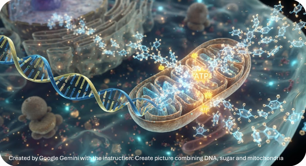
The blog examines prevalent hypotheses about cancer processes which acknowledge interactions and functions such as immune, metabolic and evolutionary pressures. In particular, the paper argues in favor of revisiting the Warburg effect and the role of mitochondrial dysfunction in cancer development. The Warburg metabolic theory proposes that impaired mitochondrial energy production may drive cells toward an ancient survival pathway characterized by increased glucose fermentation, loss of specialized cellular function, and uncontrolled or unregulated proliferation. This perspective does not reject the strong role of genetics in cancer processes but rather places DNA and RNA within a broader biological context influenced by metabolism, epigenetics, and the environment. Understanding cancer as a systemic metabolic disorder may expand prevention strategies and complement existing treatments by emphasizing lifestyle, environmental, and metabolic interventions.
Twin Studies: Genetics as Predisposition, Not Destiny
For decades, research on cancer causes and treatments has been dominated by the Somatic Mutation Theory (SMT), which proposes that cancers arise through the accumulation of DNA mutations (Weinberg, 2023; Huang et al., 2025). This SMT framework has guided the “War on Cancer” and has led to remarkable advances in targeted therapies, chemotherapy, immunotherapy, and radiation treatments. These strategies have saved countless lives, yet they primarily focus on eliminating malignant cells rather than understanding why normal cells become malignant in the first place. While a variety of mutation-focused approaches has yielded remarkable therapeutic successes, one could expect that genetically identical individuals would develop the same or similar cancers.
Decades of twin research suggests otherwise. In the landmark Nordic Twin Study of Cancer, Lichtenstein and colleagues (2000) analyzed nearly 45,000 pairs of monozygotic (identical) and dizygotic (fraternal) twins from Sweden, Denmark, and Finland. If one identical twin developed cancer, the co-twin’s probability of developing the same cancer was about 10% which is much lower than expected if inherited genes alone determined cancer risk. Heritable factors accounted for only a portion of susceptibility (approximately 27% for breast cancer, 35% for colorectal cancer, and 42% for prostate cancer). The remaining risk was attributed largely to environmental influences, lifestyle, aging, and biological processes that occur during life (Lichtenstein et al., 2000; Mucci et al., 2016; Harris et al., 2019).
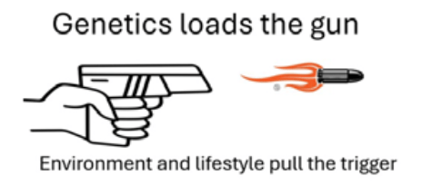
Both specific cancer investigations along with epidemiological findings across many places, people, and types of cancers suggest that genes create a predisposition, but they do not inevitably determine whether cancer develops. Instead, the cellular environment, metabolic health, environmental exposures, and epigenetic regulation strongly influence whether that predisposition is expressed. Simplistically stated, Genetics loads the gun and lifestyle and environment pulls the trigger (Peper et al., 2026).
The success of cancer treatments based on current oncology theories means that the Somatic Mutation Theory (SMT) provides a partial explanation of cancer processes. Extending the SMT of carcinogenesis to include Warburg-like theories has led to increasing evidence which suggests that metabolic dysfunction, mitochondrial injury, chronic inflammation, immune dysregulation, and environmental exposures all contribute to carcinogenesis. A useful framing of cancer risk related to inherent or heritable factors in comparison to non-intrinsic or non-genetic factors has been described by others (Brennan & Davey-Smith, 2022; Karras et al., 2024; Rahman et al., 2018; Wu et al., 2018).
For example, Wu et al., (2018) suggest that it is not possible to modify ‘random errors in DNA replication’ as an intrinsic risk factor, it is possible to partially modify endogenous risk factors related to inflammation or hormone production or fully mediate or modify directly and indirectly moderate non-genetic exogenous risk factors associated with toxic carcinogenic exposures that include exposures to some products from carcinogenic interactions with bacterial, viral, fungal and parasitic products, tobacco products, ultra-processed food and beverage products and chemicals (e.g., preservatives, pesticides, contaminants, dyes, non-nutritive chemicals such as sweeteners), industrial pollutant and chemical products (e.g. endocrine disrupting ‘forever’ chemicals such as in plastics), and lifestyle moderators such as lack of sleep, exercise, relaxation from mental ruminations and strain. A simplified model pathway would be: Risk of cancer is based on intrinsic genetic factors plus non-intrinsic endogenous and exogenous ‘epigenetic’ factors moderated by individual adaptive capacity to reduce endogenous or mitigate exogenous non-intrinsic factors.
Rather than competing explanations, mechanisms of cancerous disease processes may interact to determine whether genetically susceptible cells remain healthy or progress toward malignancy. These factors may explain that the overall cancer mortality has declined substantially over the past several decades and is predominantly due to the decreased tobacco use, earlier detection, and improvements in treatment. At the same time, the incidence of several cancers, including colorectal and breast cancer, has increased among younger adults (Siegel et al., 2025; American Cancer Society, 2024; Ugai et al., 2022; Lee, 2026). These trends suggest that additional biological and environmental factors deserve closer examination. Beyond regulating exposure to lifestyle factors such as tobacco products as a mediator of reduced cancer mortality, theories by Warburg and colleagues have pointed to metabolic regulation, including regulation of sugars, as a mediator in cancer mortality.
The Metabolic Origin: Revisiting the Warburg Effect
An alternative framework proposes that cancer is fundamentally a disorder of cellular metabolism. This concept dates back to Otto Warburg, who received the 1931 Nobel Prize in Physiology or Medicine for discovering that cancer cells exhibit a distinctive pattern of energy metabolism (Bononi et al., 2022; Otto, 2016; Nobel Prize Outreach, 2026). He showed that cancer cells rely predominantly on glucose fermentation, an anerobic process for making energy without oxygen (e.g. glucose splits into pyruvate and makes two adenosine triphosphate molecules) which is much less efficient than making energy through oxidative phosphorylation, even when oxygen is abundant in a cancer cell. The vast majority of cancerous cell growth depends on a less efficient glucose metabolism form of energy (Kim, 2017; Warburg, Negelein & Posener, 1924). The Warburg effect has been recognized as a hallmark of cancer for nearly a century (Seyfried & Chinopoulos, 2021).
Non-cancerous healthy cells mainly generate energy by producing ATP through oxidative phosphorylation within the mitochondria. Healthy cells are metabolically flexible and can generate energy from both glucose and free fatty acid oxidation (FAO). During periods of fasting, exercise, or carbohydrate restriction, triglycerides stored in adipose tissue are broken down into free fatty acids and glycerol (Eberle, 2013). The liver also converts free fatty acids into ketone bodies, which provide an efficient alternative fuel for the brain and many other tissues when glucose availability is reduced (Wakil & Abu-Elheiga, 2009; Edwards & Mohiuddin, 2023).
When mitochondrial respiration is chronically impaired by environmental toxins, oxidative stress, metabolic dysfunction, or other factors associated with carcinogenesis, cells may shift from efficient oxidative phosphorylation to a more primitive, glycolytic mode of energy production that can generate ATP in the absence of oxygen. Mitochondrial function can decline as people age when accumulation of reactive oxygen species which can directly harm mitochondrial DNA (Bondy, 2024; Cui et al., 2012; Madamanchi & Runge, 2007). In addition, mitochondrial function can also decline due to the indirect moderating effects of lifestyle choices (Caturano et al., 2025; Lemos et al., 2023; San-Millán, 2023).
As described by Miwa et al., (2022), when mitochondrial function declines, mitochondrial dysfunction can contribute to cellular senescence which is the hallmarks of aging. As a response healthy cells can progressively relinquishes their specialized role within the tissue and adopts characteristics of a more ancestral, less differentiated state (Zhang et al., 2025).
Zhang et al. (2025) suggests a few mechanisms which explain the shift of healthy cells away from specialized states towards a less differentiated state, such as shifts in the tricarboxylic acid cycle (TCA) which rely on healthy mitochondria to produce cofactors such as acetyl-CoA and positively charged Nicotinamide Adenine Dinucleotide (NAD+), along with drops in chromatin, inhibiting DNA and histone demthylases (locking chromatin in a hyper-methylated state, and an upregulation of glycolysis. Additionally, cells ‘sense’ when mitochondria have low energy production such as a high ratio of adenosine mono-phosphate or di-phosphate (AMP/ADP) to adenosine tri-phosphate (ATP), which then leads to the AMP-activated protein kinase (AMP-K) pathway (Mihaylova & Shaw, 2011). Finally, hypoxia-inducing factor (HIF) signaling is how cells ‘sense’ and adapt to changes in available oxygen, and HIF binds to DNA, activating specific genes (Huang et al., 2023). The Warburg theory suggests that when mitochondrial dysfunction directly or indirectly reduces oxidative phosphorylation, the cells shift energy production from the mitochondria to cytoplasmic glycolysis which is a hallmark of unicellular ‘ancestral’ organisms or more embryonic or stem-like cellular activity (Suomalainen, & Nunnari, 2024; Zong et al., 2024).
It is as if your cells suddenly forget they’re part of a team. Instead of working together like citizens in a well-organized society, they revert to their most primitive programming; the “me first” mentality of our single-celled ancestors. This is what happens in cancer where sophisticated multicellular cooperation gets hijacked by an ancient survival script buried deep in our biological software. Sonnenschein and Soto (1999) describe this process in their book The Society of Cells (1999), “the default state of cells is proliferation.” In other words, multiplication isn’t some aberrant behavior; it’s actually the factory setting when single cells began. Every cell carries this “go forth and multiply” command like prehistoric programming code.
In healthy tissue, cells have learned to override this ancient impulse. They’ve evolved sophisticated “stop” signals, quality control mechanisms, and cooperative protocols that keep the peace. Cancer occurs when these civilized controls break down, and cells revert to their evolutionary factory settings—endless growth, damn the consequences.
It’s as if your cells suddenly decide to ignore millions of years of evolutionary teamwork and go back to playing by the rules that worked when life was just lone microbes floating in primordial soup.
Under Survival Threat, Organisms Revert to Their Oldest Survival Mechanisms
The same regression process can be observed in human beings. When children experience overwhelming stress, they often revert to earlier developmental behaviors, such as becoming incontinent or seeking protection by hiding behind a parent. Similarly, when adults perceive an immediate threat to survival, the stress response is not always fight or flight. Instead, the body may enter a freeze response characterized by profound immobilization.
This freeze response represents an ancient survival strategy that is activated when the nervous system perceives extreme, life-threatening danger. In this state, heart rate slows, metabolism is reduced, and movement is inhibited through activation of the dorsal vagal complex which is the evolutionarily older branch of the vagus nerve according to polyvagal theory, (Porges, 2023). For reptiles and other primitive vertebrates, this physiological strategy can increase the likelihood of survival until the danger has passed.
Metabolic Theory in Context
A ‘metabolic perspective’ related to carcinogenicity, or related to reversion to more primitive types of behaviors, does not dismiss the importance of genetics. Rather, a metabolic perspective places genetic mutations within a broader biological framework in which metabolism, mitochondrial integrity, immune surveillance, environmental exposures, and epigenetic regulation interact to determine whether a cell remains healthy, or progresses toward malignancy. From this perspective, cancer represents more than the accumulation of genetic mutations. It reflects a regression to an ancient cellular survival program in which energy production and reproduction, rather than specialized function and cooperation with neighboring cells, becomes the overriding priority. Genetic mutations may therefore be viewed not only as the initiating a set of complex interactions that reflect causes of cancer but also as downstream consequences of chronic metabolic dysfunction and mitochondrial damage.
If cancer represents the activation of an evolutionarily ancient cellular survival program triggered by metabolic dysfunction, then interventions that improve metabolic health can reduce risk associated with carcinogenesis which complement conventional treatment. As Dang (2012) points out, “excessive caloric intake is associated with an increased risk for cancers, while caloric restriction is protective, perhaps through clearance of mitochondria or mitophagy, thereby reducing oxidative stress.”
One practical implication is for people to follow often repeated advice: decrease the availability of rapidly absorbed carbohydrates by minimizing the consumption of sugar-sweetened beverages, refined starches, and ultra-processed foods while emphasizing whole, nutrient-dense foods. Broadly stated, such dietary changes in sugar intake can improve metabolic resilience, reduce chronic inflammation, and create a physiological environment that is less favorable for the metabolic adaptations observed in many cancers. Although additional clinical research continues to provide evidence supporting the effectiveness of ‘lifestyle’ and epigenetic strategies across different cancer types, the metabolic perspective offers a compelling framework for prevention and adjunctive therapy.
The metabolic theory of cancer based upon the Warburg effect offers hope in treatment by reducing glucose availability may help slow the growth of some cancers. The process is described clearly by Professor Thomas Seyfried of Boston College (Seyfried et al., 2014; Seyfried et al., 2021). His metabolic theory remains an active area of research, however is not the current consensus view on cancer biology. Consider watching the compelling overview of this metabolic theory presented by Professor Thomas Seyfried’s in his YouTube lecture, Cancer as a Metabolic Disease,
What can you do to reduce cancer risk and support healing
The metabolic theory of cancer presented in this paper suggests behavioral and lifestyle strategies to reduce cancer risk and slow or prevent cancer growth. Simply stated, reduce excessive glucose availability which becomes the main energy source for cancer cells. Clinical improvement may be possible if cancer cells are put on a diet (Stetka, 2016). Changing diet is one component to optimize what you can do to reduce cancer risk, support our immune system to promote healing and optimize health although the outcome is not totally in our hands. Implement the following environmental and lifestyle behaviors promote health and healing.
Reduce and eliminate ultra-processed foods, simple carbohydrates and sugar. These increase the risk of cancers by 20 to 50 percent. Sadly, many patients undergoing chemo and radiation therapy and have difficulty with swallowing, are recommended to drink oral caloric rich supplements (such as Ensure Plus or Boost Very High Calorie) that are specially formulated to provide dense calories and protein in a small volume. They contain between 15 to 22 grams of sugar (1 to 2 tablespoons of sugar) in an 8 ounce serving– the sugar which paradoxically encourages cancer growth.
Reduce and eliminate exposure to endocrine disruptors. These are chemicals that can mimic, block, or interfere with the body’s hormones, due to higher estrogen levels (in milk and beef, for example) and pesticides (some of which act as estrogen mimics). This means eliminating as much as possible all plastics and eat mainly organic foods that do not contain herbicides or pesticides. If possible, eat organic foods.
Reduce air and water pollution that are known factors to cause cancers. This means use air and water filters at home since more than 50% of drinking water in the United States contain cariogenic substances and air pollution from car or fires are harmful.
Increase physical activity. Movement/exercise is important because that reduces lymphatic circulation and blood flow, impacting the immune system in the long term.
For detailed information and recommendations what you can use to reduce cancer risk and optimize health, see our book, Cancer Reconsidered-Why Environment, Lifestyle and Immunity Matter more than we thought.
Additional relevant blogs
References
American Cancer Society. (2024). Cancer facts & figures 2024. https://doi.org/10.3322/caac.21820
Bondy, S. C. (2024). Mitochondrial dysfunction as the major basis of brain aging. Biomolecules, 14(4), 402. https://doi.org/10.3390/biom14040402
Bononi, G., Masoni, S., Di Bussolo, V., Tuccinardi, T., Granchi, C., & Minutolo, F. (2022). Historical perspective of tumor glycolysis: A century with Otto Warburg. Seminars in Cancer Biology, 86, 325–333. https://doi.org/10.1016/j.semcancer.2022.07.003
Brennan, P., & Davey-Smith, G. (2022). Identifying novel causes of cancers to enhance cancer prevention: New strategies are needed. Journal of the National Cancer Institute, 114(3), 353–360. https://doi.org/10.1093/jnci/djab204
Caturano, A., Rocco, M., Tagliaferri, G., Piacevole, A., Nilo, D., Di Lorenzo, G., Iadicicco, I., Donnarumma, M., Galiero, R., Acierno, C., Sardu, C…., (2025). Oxidative stress and cardiovascular complications in type 2 diabetes: From pathophysiology to lifestyle modifications. Antioxidants, 14(1), Article 72. https://doi.org/10.3390/antiox14010072
Cui, H., Kong, Y., & Zhang, H. (2012). Oxidative stress, mitochondrial dysfunction, and aging. Journal of Signal Transduction, 2012, Article 646354. https://doi.org/10.1155/2012/646354
Dang, C. V. (2012). Links between metabolism and cancer. Genes & Development, 26(9), 877–890. https://doi.org/10.1101/gad.189365.112
Eberle, S. G. (2013). The body’s fuel sources. In Endurance sports nutrition (3rd ed., pp. 31–53). Human Kinetics.https://us.humankinetics.com/blogs/excerpt/the-bodys-fuel-sources
Edwards, M., & Mohiuddin, S. S. (2023, July 17). Biochemistry, lipolysis. In StatPearls. StatPearls Publishing. https://www.ncbi.nlm.nih.gov/books/NBK560564/
Hanahan, D. (2026). The hallmarks of cancer: 25 years guiding discovery and therapy. Cell, 189. https://www.cell.com/cell/fulltext/S0092-8674(26)00334-X
Hanahan, D., & Weinberg, R. A. (2011). Hallmarks of cancer: The next generation. Cell, 144(5), 646–674. https://doi.org/10.1016/j.cell.2011.02.013
Harris, J. R., Hjelmborg, J., Adami, H.-O., Czene, K., Mucci, L., & Kaprio, J. (2019). The Nordic Twin Study on Cancer (NorTwinCan). Twin Research and Human Genetics, 22(6), 817–823. https://doi.org/10.1017/thg.2019.71
Huang, S., Soto, A. M., & Sonnenschein, C. (2025). The end of the genetic paradigm of cancer. PLOS Biology, 23(3), Article e3003052. https://doi.org/10.1371/journal.pbio.3003052
Huang, X., Zhao, L., & Peng, R. (2023). Hypoxia-inducible factor 1 and mitochondria: An intimate connection. Biomolecules, 13(1), Article 50. https://doi.org/10.3390/biom13010050
Karras, P., Black, J. R. M., McGranahan, N., & Marine, J.-C. (2024). Decoding the interplay between genetic and non-genetic drivers of metastasis. Nature, 629(8012), 543–554. https://doi.org/10.1038/s41586-024-07302-6
King, M.-C., Marks, J. H., & Mandell, J. B. (2003). Breast and ovarian cancer risks due to inherited mutations in BRCA1 and BRCA2. Science, 302(5645), 643–646. https://doi.org/10.1126/science.1088759
Kim, S. Y. (2017). Cancer energy metabolism: Shutting power off cancer factory. Biomolecules & Therapeutics, 26(1), 39–44. https://doi.org/10.4062/biomolther.2017.184
Lee, I. (2026). The increase of early-onset colorectal cancer: New insights and emerging hypotheses. Cancer Control, 33, Article 10732748261432271. https://doi.org/10.1177/10732748261432271
Lemos, G. O., Torrinhas, R. S., & Waitzberg, D. L. (2023). Nutrients, physical activity, and mitochondrial dysfunction in the setting of metabolic syndrome. Nutrients, 15(5), Article 1217. https://doi.org/10.3390/nu15051217
Lichtenstein, P., Holm, N. V., Verkasalo, P. K., Iliadou, A., Kaprio, J., Koskenvuo, M., Pukkala, E., Skytthe, A., & Hemminki, K. (2000). Environmental and heritable factors in the causation of cancer: Analyses of cohorts of twins from Sweden, Denmark, and Finland. New England Journal of Medicine, 343(2), 78–85. https://doi.org/10.1056/NEJM200007133430201
Madamanchi, N. R., & Runge, M. S. (2007). Mitochondrial dysfunction in atherosclerosis. Circulation Research, 100(4), 460–473. https://doi.org/10.1161/01.RES.0000258450.44413
Mihaylova, M. M., & Shaw, R. J. (2011). The AMPK signalling pathway coordinates cell growth, autophagy and metabolism. Nature Cell Biology, 13, 1016–1023. https://doi.org/10.1038/ncb2329
Miwa, S., Kashyap, S., Chini, E., & von Zglinicki, T. (2022). Mitochondrial dysfunction in cell senescence and aging. Journal of Clinical Investigation, 132(13), Article e158447. https://doi.org/10.1172/JCI158447
Mucci, L. A., Hjelmborg, J. B., Harris, J. R., et al. (2016). Familial risk and heritability of cancer among twins in Nordic countries: The Nordic Twin Study of Cancer (NorTwinCan) collaboration. JAMA, 315(1), 68–78. https://doi.org/10.1001/jama.2015.17703
Mukherjee, S. (2025). The emperor of all maladies: A biography of cancer. Scribner. https://www.amazon.com/Emperor-All-Maladies-Biography-Cancer/dp/1668047039/
National Institutes of Health. (2025, April 21). Annual report to the nation: Cancer deaths continue to decline [Media advisory]. NIH News Releases. https://www.nih.gov/news-events/news-releases/annual-report-nation-cancer-deaths-continue-decline
Nobel Prize Outreach. (2026). Otto Warburg—Biographical. NobelPrize.org. Retrieved August 11, 2026, from https://www.nobelprize.org/prizes/medicine/1931/warburg/biographical/
Otto, A. M. (2016). Warburg effect(s)—A biographical sketch of Otto Warburg and his impacts on tumor metabolism. Cancer & Metabolism, 4(1), Article 5. https://doi.org/10.1186/s40170-016-0145-9
Peper, E., Gorter, R., & Faass, N. (2026). Cancer reconsidered: Why environment, lifestyle, and immunity matter more than we thought. BiofeedbackHealth/Regent Press. https://www.amazon.com/Cancer-Reconsidered-Environment-Lifestyle-Immunity/dp/1587907402
Porges, S. W. (2023). The vagal paradox: A polyvagal solution. Comprehensive Psychoneuroendocrinology, 16, Article 100200. https://doi.org/10.1016/j.cpnec.2023.100200
Rahman, M. S., Suresh, S., & Waly, M. I. (2018). Risk factors for cancer: Genetic and environment. In Bioactive Components, Diet and Medical Treatment in Cancer Prevention (pp. 1-23). Springer International Publishing. https://doi.org/10.1007/978-3-319-75693-6_1
San-Millán, I. (2023). The key role of mitochondrial function in health and disease. Antioxidants, 12(4), Article 782. https://doi.org/10.3390/antiox12040782
Seyfried, T. N., & Chinopoulos, C. (2021). Can the mitochondrial metabolic theory explain better the origin and management of cancer than can the somatic mutation theory? Metabolites, 11(9), Article 572. https://doi.org/10.3390/metabo11090572
Seyfried, T. N., Flores, R. E., Poff, A. M., & D’Agostino, D. P. (2014). Cancer as a metabolic disease: Implications for novel therapeutics. Carcinogenesis, 35(3), 515–527. https://doi.org/10.1093/carcin/bgt480
Seyfried, T. N., Lee, D. C., Duraj, T., et al. (2025). The Warburg hypothesis and the emergence of the mitochondrial metabolic theory of cancer. Journal of Bioenergetics and Biomembranes, 57, 57–83. https://doi.org/10.1007/s10863-025-10059-w
Siegel, R. L., Kratzer, T. B., Giaquinto, A. N., Sung, H., & Jemal, A. (2025). Cancer statistics, 2025. CA: A Cancer Journal for Clinicians, 75(1), 10–45. https://doi.org/10.3322/caac.21871
Sonnenschein, C., & Soto, A. M. (1999). The society of cells: Cancer and control of cell proliferation. BIOS Scientific Publishers. https://www.amazon.com/Society-Cells-Carlos-Sonnenschein/dp/1859962769
Stetka, B. (2016, March 5). Fighting cancer by putting tumor cells on a diet. NPR. https://www.npr.org/sections/health-shots/2016/03/05/468285545/fighting-cancer-by-putting-tumor-cells-on-a-diet
Suomalainen, A., & Nunnari, J. (2024). Mitochondria at the crossroads of health and disease. Cell, 187(11), 2601–2627. https://doi.org/10.1016/j.cell.2024.04.037
Ugai, T., Sasamoto, N., Lee, H. Y., Ando, M., Song, M., Tamimi, R. M., … Ogino, S. (2022). Is early-onset cancer an emerging global epidemic? Current evidence and future implications. Nature Reviews Clinical Oncology, 19(10), 656–673. https://doi.org/10.1038/s41571-022-00672-8
Wakil, S. J., & Abu-Elheiga, L. A. (2009). Fatty acid metabolism: Target for metabolic syndrome. Journal of Lipid Research, 50(Suppl.), S138–S143. https://doi.org/10.1194/jlr.R800079-JLR200
Warburg, O., Negelein, E. & Posener, K. Versuche an Überlebendem Carcinomgewebe. Klin Wochenschr 3, 1062–1064 (1924). https://doi.org/10.1007/BF01736087
Weinberg, R. A. (2023). The biology of cancer. W. W. Norton & Company. https://www.amazon.com/Biology-Cancer-Robert-Weinberg/dp/0393887650
Wu, S., Zhu, W., Thompson, P., & Hannun, Y. A. (2018). Evaluating intrinsic and non-intrinsic cancer risk factors. Nature Communications, 9(1), Article 3490. https://doi.org/10.1038/s41467-018-05467-z
Zhang, Y., Tang, J., Jiang, C., Yi, H., Guang, S., Yin, G., & Wang, M. (2025). Metabolic reprogramming in cancer and senescence. MedComm, 6(3), Article e70055. https://doi.org/10.1002/mco2.70055
Zong, Y., Li, H., Liao, P., et al. (2024). Mitochondrial dysfunction: Mechanisms and advances in therapy. Signal Transduction and Targeted Therapy, 9, 124. https://doi.org/10.1038/s41392-024-01839-8
Edited with the help of ChatGPT
Will the stock market bubble collapse? Will human fertility decrease to zero?
Posted: July 13, 2026 Filed under: behavior, cancer, Evolutionary perspective, healing, health, lifestyle, Nutrition/diet | Tags: books, cycles, economics, fertility, financial bubble, sperm count, stocks, technology Leave a comment
“Those who cannot remember the past are condemned to repeat it.” – George Santayana (Santayana, 1905)
These two questions may seem totally unrelated, yet they are deeply connected if one takes a long historic view and hypothesizes that present-day trends will continue.
When I drive or commute, I very much enjoy listening to podcasts. They offer an in-depth, nuanced discussions that are missing from today’s soundbite culture. These deep-dive podcasts offer a vital antidote, providing the nuanced, extended discussions necessary for true cognitive synthesis. One of my favorite podcasts is The Diary of a CEO with Steven Bartlett. I found the recent episode, “Billionaire’s WARNING: I’m SELLING. The Crash Is Already Here!”, very eye opening.
The interview was with billionaire investor Jeremy Grantham, the co-founder and long-term investment strategist of GMO. He is also the chairman of the Grantham Foundation for the Preservation of the Environment, and co-author of “The Making of a Permabear: The Perils of Long-term Investing in a Short-term World.”
While economics and financial markets fall outside my primary line of research, I deeply appreciated his long-term perspective—an analysis I find to be entirely spot-on. The actual content offered a masterclass in pattern recognition, forcing me to sit back and critically analyze the macroeconomic shifts happening around us rather than just reacting to the daily noise of the market. He persuaded me that the present AI-driven financial bubble will burst; we simply do not know exactly when. I concur with him that it will occur sooner rather than later. Personally, I would not be surprised if it occurred within the next two years. We must simply be prepared to deal with more challenging times.
What surprised and fascinated me even more were the last 40 minutes of the podcast. Grantham shifted entirely away from the financial markets and applied that same rigorous, analytical thinking to what is truly driving the modern “baby bust” (Dilmaghani et al., 2024).
The scientific data is clear: human fertility—both male and female—has been decreasing each decade. This is starkly indicated by a 50% drop in male sperm counts and a rising number of couples struggling to conceive (Ravitsky & Kimmins, 2019; Mann et al., 2020; Inam, 2025). The culprits as for the increase in cancer rates? Ubiquitous modern disruptions like microplastics, pesticides, and pervasive environmental toxins (NIOSH, 2023; Doroftei et al., 2025; Brander et al., 2026).
Although the prognosis can look bleak, Grantham doesn’t just sound the alarm; he offers realistic strategies. He outlines how to protect yourself from the impending financial bubble collapse, as well as what you and your community can do to minimize toxic exposure and enhance fertility.
I smiled as I listened to the podcast, because his systemic analysis and practical recommendations are very similar to what we described in our just-published book, Cancer reconsidered-Why Environment, lifestyle, and immunity matter more than we thought (Peper et al., 2026).
To prepare yourself for the financial future and optimize your health and fertility, listen to Grantham’s insights on the podcast episode, “Billionaire’s WARNING: I’m SELLING. The Crash Is Already Here!”,
See the following blogs for more detailed information:
References
Brander, S.M., Swan, S.H., Mehinto, A.C., & et al. (2026). Impacts of environmental stressors on fertility and fecundity across taxa, with implications for planetary health. npj Emerging Contaminants, 2, Article 12. https://doi.org/10.1038/s44454-026-00032-6
Dilmaghani, D., Ainsworth, A. J., Nath, K. A., & Garovic, V. D. (2024). Decreasing fertility rate in the United States: Demographics, challenges, and consequences. Mayo Clinic Proceedings, 99(11), 1693–1697. https://doi.org/10.1016/j.mayocp.2024.09.004
Doroftei, B., Savuca, A., Cretu, A.-M., Maftei, R., Anton, N., Ilea, C., Doroftei, M., & Puha, B. (2025). Microplastics and human fertility: A comprehensive review of their presence in human samples and reproductive implication. Ecotoxicology and Environmental Safety, 303, Article 118939. https://doi.org/10.1016/j.ecoenv.2025.118939
Inam, Ö. (2025). Impact of microplastics on female reproductive health: Insights from animal and human experimental studies: A systematic review. Archives of Gynecology and Obstetrics, 312(1), 77–92. https://doi.org/10.1007/s00404-024-07929
Mann, U., Shiff, B., & Patel, P. (2020). Reasons for worldwide decline in male fertility. Current Opinion in Urology, 30(3), 296–301. https://doi.org/10.1097/MOU.0000000000000745
NIOSH. (2023, December 15). About pesticides and reproductive health. National Institute for Occupational Safety and Health Centers for Disease Control and Prevention. https://www.cdc.gov/niosh/reproductive-health/prevention/pesticides.html
Peper, E. Gorter, R. & Faass, N. (2026). Cancer Reconsidered: Why Environment, Lifestyle, and Immunity Matter More Than We Thought. BiofeedbackHealth/Regent Press. https://www.amazon.com/Cancer-Reconsidered-Environment-Lifestyle-Immunity/dp/1587907402/
Ravitsky, V. & Kimmins, S. (2019). The forgotten men: rising rates of male infertility urgently require new approaches for its prevention, diagnosis and treatment. Biol Reprod. 101(5), 872-874. https://doi.org/10.1093/biolre/ioz161
Santayana, G. (1905). The Life of Reason: Vol. 1. Reason in Common Sense. Charles Scribner’s Sons. p. 284. https://www.amazon.com/Reason-Phases-Progress-Introduction-Common/dp/B01DF937WU
Can Your Daily Choices Influence Cancer Risk and Recovery?
Posted: June 27, 2026 Filed under: behavior, Breathing/respiration, cancer, emotions, healing, health, lifestyle, Nutrition/diet, Pain/discomfort, self-healing, stress management, Uncategorized | Tags: diet, endocrine disregulators, environmental toxins, glucose monitors, health, Immune function, mental-health, nutrition, pollution, Quality of life, regeneration, resilience, social support, wellness 1 CommentWhat if the way you eat, move, sleep, manage stress, and connect with others could influence your body’s ability to prevent disease and support healing?
Most of us have been taught to think of cancer primarily as a genetic disease. Yet an expanding body of scientific research tells a more hopeful story: while genes matter, they are only part of the picture. Our environment, lifestyle, immune system, and even the quality of our relationships can profoundly influence health.
These are the questions explored in the newly published book Cancer Reconsidered: Why Environment, Lifestyle, and Immunity Matter More than We Thought, by Erik Peper, Robert Gorter, and Nancy Faass.
Written for people living with cancer, their families, healthcare professionals, and anyone interested in optimizing health, the book translates decades of scientific research into practical, evidence-based strategies that readers can use in everyday life.
Rather than viewing cancer through a single lens, Cancer Reconsidered brings together insights from conventional medicine with evidence-based complementary approaches. The authors explore how nutrition, physical activity, stress, sleep, environmental exposures, immune function, and social support interact to influence both cancer risk and the body’s remarkable capacity for repair and resilience.
One of the book’s central messages is both simple and empowering: although we cannot change our genes, we can often change the conditions in which our genes are expressed. Daily choices matter. Healthy habits can strengthen the body’s natural defenses, reduce inflammation, support immune function, and improve quality of life.
A particularly practical chapter explores blood sugar regulation and metabolism. Using continuous glucose monitors (CGMs) together with smartphone apps, readers can observe in real time how different foods, exercise, stress, and sleep affect their glucose levels. Instead of following one-size-fits-all advice, they become active investigators of their own health, discovering what works best for their unique physiology.
Throughout the book, the emphasis is not on fear, but on possibility. Scientific evidence increasingly shows that hope, meaningful social connections, regular movement, nourishing food, restorative sleep, effective stress management, and resilience are not simply “nice ideas”—they are biological factors that can significantly influence health and well-being.
Cancer Reconsidered invites readers to move beyond the question, “What causes cancer?” and instead ask, “What can I do today to create the best possible conditions for health?” It offers a thoughtful, scientifically grounded roadmap for anyone seeking to answer that question.
Cancer Reconsidered: Why Environment, Lifestyle, and Immunity Matter More than We Thought is now available on Amazon in paperback and and affordable ebook Kindle editions. https://www.amazon.com/s?k=cancer+reconsidered
Managing Blood Sugar: Lifestyle Changes for Better Health
Posted: May 25, 2026 Filed under: ADHD, attention, behavior, cancer, Evolutionary perspective, healing, health, lifestyle, Nutrition/diet, self-healing, Uncategorized | Tags: AIC, carbohydrate, continous glucose monitoring, diet, exhaustion, glucose, insulin, metabolic disease, obesity, sugar, ultra-processed foods Leave a comment
It was 4:45 p.m., and I was looking forward to swimming. I briskly walked the eight blocks from my house to the heated outdoor city pool. The pool is unusual—100 feet long instead of the standard 25 yards—and I enjoy the rhythm of swimming lap after lap.
I arrived just as the sign flipped from Closed to Open. I quickly changed into my bathing suit, locked my clothes in a metal locker, took a short shower, and jumped into the lane. The sun was still out, and only one other swimmer shared my lane. I felt energized and expected to complete my usual forty laps.
However, after about eighteen laps, my energy suddenly disappeared. There was nothing left in the tank. I swam to the side, pulled myself slowly onto the pool deck, and even slowly and unsteadily walked to the men’s locker room. I sat down on the bench feeling shaky, weak, and exhausted. This was not ordinary fatigue. After resting for several minutes, I slowly showered, dressed, and walked home with heavy, almost uncoordinated legs.
As I reflected on the experience, I remembered something similar that had happened two weeks earlier. Around 5 p.m, I had taken my son’s dog for a brisk walk. Again, I began energized, walking quickly, and then suddenly felt drained and sweaty. When I returned home, all I could do was sit down and recover.
What happened
Reflecting back, I realized that both cases I had eaten sweets—cake one time and a large chocolate chip cookie the other about two hours earlier Most likely, the rapidly absorbed sugars and refined carbohydrates caused a sharp increase in blood glucose, followed by a significant insulin response (Ludwig & Ebbeling, 2018). During exercise, my muscles then demanded additional glucose, and my blood sugar may have dropped rapidly enough to trigger symptoms of reactive hypoglycemia: shakiness, sweating, weakness, and fatigue (Morales-Brown, 2025, June 12).
The process is more complex than simply “sugar highs” and “crashes.” Carbohydrates are broken .break down into glucose during digestion, which begins in the mouth. Chewing breaks down food physically, while the enzyme amylase in saliva starts the chemical breakdown by splitting starches into sugars (Peyrot des Gachons & Breslin, 2016). This raises blood glucose levels, which stimulates insulin release from the pancreas. Insulin helps move glucose from the bloodstream into cells. In some people—especially those developing insulin resistance or prediabetes—the insulin response may overshoot, leading to a later drop in blood glucose. Exercise can amplify this effect because active muscles rapidly consume glucose for energy (American Diabetes Association, 2024).
This experience was a wake-up call for me because my hemoglobin A1C is 5.7%, the lower threshold for prediabetes. Hemoglobin A1C reflects average blood glucose levels over approximately the previous three months (American Diabetes Association, 2026).
Like many people, I enjoy and am even addicted to bread, potatoes, pastries, and sweets. Looking back, the subtle changes began during COVID. Before the pandemic, I spent much of the day teaching in person, walking across campus, moving, and interacting with students. During lockdown, I sat for hours teaching online. My physical activity during the day dramatically decreased while my eating habits did not significantly change.
When we are inactive, excess sugars and refined carbohydrates are less likely to be immediately used by muscles for fuel. Instead, repeated spikes in blood glucose and insulin can contribute over time to insulin resistance, weight gain, metabolic dysfunction, and increased inflammation (Ludwig & Ebbeling, 2018).
Although my episodes were minor, they reminded me that lifestyle patterns especially eating ultra-processed foods can increase the risk for chronic diseases such as type 2 diabetes, cardiovascular disease, obesity, and some cancers and dementia (Lane et al., 2024; Menegassi & Vinciguerra, 2025). The scientific literature strongly links obesity, insulin resistance, and type 2 diabetes with increased risk for several cancers, including colorectal and postmenopausal breast cancer (Peper et al., 2026; Scully et al., 2021: Lauby-Secretan et al., 2016). Ultra-processed foods and sugar-sweetened beverages are also associated with increased risk for obesity and metabolic disease; moreover, cancer survivors who consume higher amounts of ultra processed foods face a significantly increased risk of both all-cause and cancer-specific mortality (Hall et al., 2019; Bonaccio et al., 2026). However, cancer is multifactorial, and no single food alone “causes” cancer. Rather, long-term dietary patterns, inactivity, obesity, chronic inflammation, genetics, environmental exposures, sleep, and stress all interact together ( Marino et al., 2024; Dalamaga et al., 2026; Peper et al., 2026).
What to do
The encouraging news is that these processes are often reversible.
Weight, hunger, blood sugar fluctuations, and even A1C are not fixed. They can improve significantly through lifestyle changes. Research consistently shows that reducing ultra-processed foods, lowering intake of refined carbohydrates and sugary beverages, increasing fiber-rich vegetables, improving sleep, reducing stress, and exercising regularly can improve insulin sensitivity and metabolic health (Bird & Hawley, 2017; Vaezi et al., 2025; American Diabetes Association, 2024; Peper et al., 2026).
For many people, continuous glucose monitors (CGMs) can provide powerful real-time feedback (Ehrhardt & Zaghal, 2020). Seeing how specific foods affect your glucose levels can increase awareness and motivate healthier choices. Often, we do not realize how dramatically a muffin, fruit juice, or bowl of white rice may affect blood sugar until we see the data on the screen.
The goal is not perfection or rigid dieting. Instead, it is learning to observe how your body responds and gradually shifting toward foods that support stable energy, satiety, and long-term health.
Before making major dietary changes, watch the superb interview with Dr. David Unwin, a British physician known for his work using lower-carbohydrate dietary approaches to help patients improve type 2 diabetes and metabolic health. His clinical work demonstrates that many patients can significantly improve blood sugar control and sometimes reduce medications through lifestyle changes (Unwin et al., 2020). The video, The Sugar Doctor’s Warning: The “Healthy” Foods Quietly Destroying Your Body! – Dr. David Unwin, is from the podcast, The Diary of a CEO with Steven Bartlett.
The Link Between Diet, Lifestyle, and Cancer Risk: Steps You Can Take
Read the new book, Cancer Reconsidered: Why Environment, Lifestyle, and Immunity Matter More than We Thought, by Erik Peper, Robert Gorter, and Nancy Faass. It explore the many of the lifestyle factors that can increase cancer risk—or help protect against it. The book brings together an extraordinary range of scientific research to illuminate how everyday habits and modern lifestyles influence cancer risk and healing. Drawing from both conventional medicine and integrative approaches, the authors thoughtfully examine the many factors involved in cancer causation while offering hopeful, evidence-based strategies for supporting recovery and restoring health.
What makes this book especially compelling is that it goes far beyond reviewing the science. It translates research into practical, everyday actions people can use to support healing and improve quality of life. At its heart is lifestyle medicine—the recognition that stress management, hope, physical activity, nourishing foods, supportive relationships, community, and resilience during times of crisis profoundly affect health and well-being. The book also offers a detailed and highly practical discussion of sugar metabolism and explains how continuous glucose monitoring sensors (CGMS) with the smartphone app can help people directly observe how specific foods and daily habits influence their blood sugar levels. Instead of relying on abstract nutrition advice, readers learn how to become active investigators of their own health.
Throughout the book, the message is empowering: our daily habits, social connections, attitudes, and environment may influence health as much as—if not more than—genetics alone. The book is available from Amazon: https://www.amazon.com/Cancer-Reconsidered-Environment-Lifestyle-Immunity/dp/1587907402
Listen to the in-depth discussion of this blog created with Google NotebookLM
Addition relevant blogs
References
American Diabetes Association. (2026). Blood glucose and exercise. American Diabetes Association. Retrieved May 24, 2026, from https://diabetes.org/health-wellness/fitness/blood-glucose-and-exercise
American Diabetes Association. (2024). Standards of care in diabetes—2024. Diabetes Care, 47(Suppl. 1). https://diabetesjournals.org/care/issue/47/Supplement_1
Bonaccio, M., Di Castelnuovo, A., Costanzo, S., Ruggiero, E., Esposito, S., Panzera, T., Di Costanzo, G., De Curtis, A., Magnacca, S., Cerletti, C., Donati, M. B., de Gaetano, G., & Iacoviello, L., for the Moli-sani Study Group. (2026). Ultra-processed food and mortality among long-term cancer survivors from the Moli-sani Study: Prospective findings and analysis of biological pathways. Cancer Epidemiology, Biomarkers & Prevention, 35(4), 664–674. https://doi.org/10.1158/1055-9965.EPI-25-0808
Dalamaga, M., Rozani, S., & Petropoulou, D. (2026). Why is colorectal cancer occurring earlier? Metabolic dysfunction, underrecognized carcinogens, and emerging controversies. Current Obesity Reports, 15(1), 24. https://doi.org/10.1007/s13679-026-00700-z
Ehrhardt, N., & Al Zaghal, E. (2020). Continuous glucose monitoring as a behavior modification tool. Clinical Diabetes, 38(2), 126–131. https://doi.org/10.2337/cd19-0037
Hall, K. D., Ayuketah, A., Brychta, R., Cai, H., Cassimatis, T., Chen, K. Y., Chung, S. T., Costa, E., Courville, A., Darcey, V., Fletcher, L. A., Forde, C. G., Gharib, A. M., Guo, J., Howard, R., Joseph, P. V., McGehee, S., Ouwerkerk, R., Raisinger, K., … Zhou, M. (2019). Ultra-processed diets cause excess calorie intake and weight gain: An inpatient randomized controlled trial. Cell Metabolism, 30(1), 67–77. https://doi.org/10.1016/j.cmet.2019.05.008
Lane, M. M., Gamage, E., Du, S., Ashtree, D. N., McGuinness, A. J., Gauci, S., Baker, P., Lawrence, M., Rebholz, C. M., Srour, B., Touvier, M., Jacka, F. N., O’Neil, A., Segasby, T., & Marx, W. (2024). Ultra-processed food exposure and adverse health outcomes: Umbrella review of epidemiological meta-analyses. BMJ, 384, e077310. https://doi.org/10.1136/bmj-2023-077310
Lauby-Secretan, B., Scoccianti, C., Loomis, D., Grosse, Y., Bianchini, F., & Straif, K. (2016). Body fatness and cancer—Viewpoint of the IARC Working Group. New England Journal of Medicine, 375(8), 794–798. https://doi.org/10.1056/NEJMsr1606602
Ludwig, D. S., & Ebbeling, C. B. (2018). The carbohydrate-insulin model of obesity: Beyond “calories in, calories out.” JAMA Internal Medicine, 178(8), 1098–1103. https://doi.org/10.1001/jamainternmed.2018.2933
Marino, P., Mininni, M., Deiana, G., Marino, G., Divella, R., Bochicchio, I., Giuliano, A., Lapadula, S., Lettini, A. R., & Sanseverino, F. (2024). Healthy lifestyle and cancer risk: Modifiable risk factors to prevent cancer. Nutrients, 16(6), 800. https://doi.org/10.3390/nu16060800
Menegassi, B., & Vinciguerra, M. (2025). Ultraprocessed food and risk of cancer: Mechanistic pathways and public health implications. Cancers, 17(13), 2064. https://doi.org/10.3390/cancers17132064
Morales-Brown, P. (2025, June 12). What is reactive hypglycemia. Medical News Today. Accessed May 24, 2026. https://www.medicalnewstoday.com/articles/reactive-hypoglycemia
Peper, E., Gorter, R., & Faass, N. (2026). Cancer reconsidered: Why environment, lifestyle, and immunity matter more than we thought. Biofeedback Health. https://www.amazon.com/Cancer-Reconsidered-Environment-Lifestyle-Immunity/dp/1587907402
Peyrot des Gachons, C., & Breslin, P. A. S. (2016). Salivary amylase: Digestion and metabolic syndrome. Current Diabetes Reports, 16, 102. https://doi.org/10.1007/s11892-016-0794-7
Scully, T., Ettela, A., LeRoith, D., & Gallagher, E. J. (2021). Obesity, type 2 diabetes, and cancer risk. Frontiers in Oncology, 10, 615375. https://doi.org/10.3389/fonc.2020.615375
Unwin, D., Khalid, A. A., Unwin, J., Crocombe, D., Delon, C., Martyn, K., Hasan, M., & Tobin, S. D. (2020). Insights from a general practice service evaluation supporting a lower carbohydrate diet in patients with type 2 diabetes mellitus and prediabetes: A secondary analysis of routine clinic data including HbA1c, weight and prescribing over 6 years. BMJ Nutrition, Prevention & Health, 3(2), 285–294. https://doi.org/10.1136/bmjnph-2020-000072
Vaezi, S., Freeling, J. L., de Vargas, B. O., Weidauer, L., Shoemaker, M. E., Sanders, W. M., & Dey, M. (2025). Impacts of minimally-processed omnivorous vs lacto-ovo-vegetarian diets on insulin sensitivity, lipid profile, and adiposity in older adults: Secondary findings from a randomized crossover feeding trial. Clinical Nutrition, 55, 90–103. https://doi.org/10.1016/j.clnu.2025.10.010
Improve fertility and decrease cancer risk: THE PLASTIC DETOX
Posted: May 7, 2026 Filed under: behavior, cancer, health, Uncategorized | Tags: bisphenols, BPA, endocrine disruptors, environment, environmental pollution, fertility, health, microplastics, plastics, polution, pregnancy, sperm, sustainability, wellness 3 CommentsAre you concerned about your or your children’s future fertility, or do you want to get pregnant?
Do you want to reduce your cancer risk?
In 1962, Rachel Carson published the seminal, groundbreaking book Silent Spring, which brought public awareness to the harmful effects of environmental pollution (Carson, 1962). In many cases, public awareness of environmental pollution has been driven by observations and research involving people living near or on toxic waste sites, such as the infamous Love Canal in Niagara Falls, New York. Residents living in this area experienced a significantly higher risk of developing bladder, kidney, and other cancers due to contaminated water and soil (Gensburg et al., 2009).
Equally important is the impact of environmental pollution, including ongoing exposure to plastics, on health and embryological development. Many studies report that downstream pollution from agriculture, chemical plants, and sewage causes significant harm, ranging from limb deformities in amphibians (Taylor et al., 2005) to disruptions in human reproductive health. For example, microplastics have been shown to impair ovarian function, decrease fertility rates, and disrupt hormone levels in female subjects and their offspring’s health (Inam, 2025).
Research findings suggest that many human-made chemicals reduce fertility and increase cancer risk. Many chemicals in plastics (e.g., bisphenols (BPA, BPS, BPF), phthalates, flame retardants, and nonylphenol) are endocrine disruptors contributing to reduced fertility and can act as carcinogenic initiators or promoters (NIH, 2026). Ongoing exposure to plastics is one of several factors that may contribute to declines in fertility and earlier onset of cancers such as breast and prostate cancer in younger populations.
The recently released Netflix documentary The Plastic Detox offers an eye-opening exploration of the hidden dangers of the chemicals in plastics in our homes and daily lives and how it may impact fertility. It highlights concerns ranging from hormone disruption—which may contribute to declining fertility worldwide—to increasing rates of cancer and earlier occurrences of heart attack and stroke.
In the documentary, Professor Shanna H. Swan, a research scientist at the Icahn School of Medicine at Mount Sinai in New York City, explains how microplastics and their associated chemicals may affect our health—and what steps we can take to reduce our exposure. Although the documentary is not a randomized controlled trial and it doesn’t disentangle the possible placebo effects that arise when people shift their beliefs about the cause of infertility (for example, “it’s not my fault; it’s the plastics”), its central message is valid. From a psychophysiological perspective, how we interpret the causes of our health challenges can shape both our stress responses and our sense of agency.
Even with its scientific limitations, the film points toward an important concern: that environmental exposures, including plastics, play an important role in reproductive health. I also wonder whether some of the sharp “scientific critiques” of the documentary reflect more than scientific caution alone. History reminds us that industries whose profits are threatened have often worked to amplify uncertainty and delay regulation as the tobacco industry famously did for decades despite mounting evidence of harm (Maani et al., 2022; Oreskes & Conway, 2010).
I strongly recommend watching the documentary and taking steps to reduce exposure to plastics and other environmental toxins (such as glyphosate and the chemicals in air and water pollution) to support your health and that of your children. Watch it on Netflix: https://www.netflix.com/title/82074244
Listen to the in-depth discussion of this blog created with Google NotebookLM
References
Carson, R. (1962). Silent Spring. Houghton Mifflin Co. https://www.amazon.com/Silent-Spring-Rachel-Carson/dp/0395075068
Gensburg, L. J., Pantea, C., Fitzgerald, E., Stark, A., Hwang, S. A., & Kim, N. (2009). Mortality among former Love Canal residents. Environmental Health Perspectives, 117(2), 209–216. https://doi.org/10.1289/ehp.11350
Inam, Ö. (2025). Impact of microplastics on female reproductive health: Insights from animal and human experimental studies: A systematic review. Archives of Gynecology and Obstetrics, 312(1), 77–92. https://doi.org/10.1007/s00404-024-07929
Maani, N., van Schalkwyk, M. C. I., Filippidis, F. T., Knai, C., & Petticrew, M. (2022). Manufacturing doubt: Assessing the effects of independent vs. industry-sponsored messaging about the harms of fossil fuels, smoking, alcohol, and sugar-sweetened beverages. SSM – Population Health, 17, 101009. https://doi.org/10.1016/j.ssmph.2021.101009
NIH. (2026). Endocrine disruptors. National Institute of Environmental Health Sciences. Retrieved May 7, 2026, from https://www.niehs.nih.gov/health/topics/agents/endocrine/index.cfm
Oreskes, N., & Conway, E. M. (2010). Merchants of doubt: How a handful of scientists obscured the truth on issues from tobacco smoke to global warming. Bloomsbury Press. https://www.amazon.com/Merchants-Doubt-Handful-Scientists-Obscured/dp/1608193942/
Taylor, B., Skelly, D., Demarchis, L. K., Slade, M. D., Galusha, D., & Rabinowitz, P. M. (2005). Proximity to pollution sources and risk of amphibian limb malformation. Environmental Health Perspectives, 113(11), 1497–1501. https://doi.org/10.1289/ehp.7585
Tell Me Where It Hurts
Posted: April 24, 2026 Filed under: attention, behavior, biofeedback, Breathing/respiration, cancer, education, emotions, healing, health, Neck and shoulder discomfort, Pain/discomfort, self-healing, surgery | Tags: health, meditation, mental-health, mindfulness, wellness 2 Comments
In my biofeedback practice, I’ve repeatedly seen that healing rarely comes from a single technique. It emerges from observation, integration and practicing and integrating skills in daily life. When clients learn to combine biofeedback with other strategies to make it their own such as slower, more coherent breathing, guided imagery, shifts in internal dialogue, and practical lifestyle changes, something important happens: the body begins to reorganize itself toward health.
Again and again, clients report meaningful changes. Stress symptoms, headaches, eye problem, neck shoulder and back pain decrease or disappear. Gastrointestinal symptoms often fade out. Anxiety loosens its grip. Asthma improves. Chronic neck, shoulder, and pelvic pain diminish. These are not isolated outcomes; they reflect a pattern. When people gain the skills to regulate their physiology and reinterpret their internal experience, decrease in symptoms often follow (see the list of articles that describe successful outcomes).
Even though many of my clients benefit, I am continually searching for strategies and approaches that can improve their health and reduce suffering and for materials that I can recommend to them.
Now when I see a client who reports pain or who takes care of someone with pain, the first thing I do is to recommend the book, Tell Me Where It Hurts, by Rachel Zoffness, PhD, a leading pain expert and psychologist. The book offers a clear, science-based framework grounded in modern neuroscience, yet conveyed through compelling human stories. Her work aligns closely with what we observe in biofeedback: pain is not simply a signal from injured tissue. It is an experience shaped by the interaction of body, brain, emotions, beliefs, culture and context.
She makes a crucial point that pain may begin with injury or illness, but it is always modulated by factors such as physiological state, emotional meaning, cognitive interpretation, and social and cultural influences. In other words, pain is real, but it is also dynamic and changeable.
The first step she emphasizes is education. When people understand how pain actually works, fear often decreases. From there, the task becomes identifying what amplifies pain and what reduces it and then systematically strengthening the factors and behaviors and skills that support recovery.
One striking story captures this perfectly: a construction worker jumps from a plank onto what appears to be a 7” nail, which is driven through his boot. He experiences excruciating pain and is rushed to the emergency room. Yet when the boot is removed, the nail is found to have passed cleanly between his toes—there is no tissue damage. The pain was real, but it was driven by perception and expectation. This is not an anomaly; it is a powerful illustration of how the brain constructs pain.
Equally compelling are the recovery stories. Patients with severe chronic pain that continue and got worse after failed surgeries, long-term disability, or even amputation, find relief not through more invasive procedures or medication alone, but through learning how to retrain their nervous system. In many of these cases, even opioids had failed to provide meaningful relief. What made the difference was a shift in understanding, combined with evidence-based self-regulation strategies. They are no longer abstract ideas; they are lived experiences.
The larger message is both simple and profound: pain can change. And when people are given the right framework and tools, they can actively participate in that change. For anyone living with pain, or working professionally with those who do, this book is not just informative. It is practical, empowering, and, in many cases, transformative.
This is the book to read if you have pain or care for someone with pain. It is also the book every therapist who works with people with pain should read and recommend to their clients.
Zoffness, R. (2026). Tell me where it hurts. Grand Central. https://www.amazon.com/Tell-Me-Where-Hurts-Science/dp/1538758148/
I recommend listening to the excellent podcast generated with Google NotebookLM, which thoughtfully expands upon and clarifies the underlying research.isten to the expanded podcast based on this blog and created with Google Notebook LM.
The following blog has a link to a superb podcast featuring Rachel Zoffness.
Then listen to the podcast from Dolorology (PAIN) Encore with Dr. Rachel Zoffness. https://www.alieward.com/ologies/dolorologyencore
Examples of integrated biofeedback outcomes
Peper, E. (2015). Pain as a Contextual Experience. Townsend Letter-The Examiner of Alternative Medicine, 388, 63-66. https://townsendletter.com/peper-contextual-pain-09-11-15/
Peper, E., Booiman, A. & Harvey, R. (2025a). Pain-There is Hope. Biofeedback, 53(1), 1-9. http://doi.org/10.5298/1081-5937-53.01.16 Also, republished in Townsend Letter-Innovative Health Perspectives. https://townsendletter.com/pain-there-is-hope/
Peper, E., Chen, S., Heinz, N. & Harvey, R. (2023). Hope for menstrual cramps (dysmenorrhea) with breathing. Biofeedback,. 51(2), 44–51. https://doi.org/10.5298/1081-5937-51.2.04
Peper, E. & Cohen, T. (2017). Inhale to breathe away pelvic floor pain and enjoy intercourse. Biofeedback, 45(1), 21–24. https://doi.org/10.5298/1081-5937-45.1.04
Peper, E., Cosby, J., & Almendras, M. (2022a). Healing chronic back pain. NeuroRegulation, 9(3), 164–172. https://doi.org/10.15540/nr.9.3.164
Peper, E., Covell, A., & Matzembacker, N. (2021). How a chronic headache condition became resolved with one session of breathing and posture coaching. NeuroRegulation, 8(4), 194–197. https://doi.org/10.15540/nr.8.4.194
Peper, E. & Harvey, R. (2008). From technostress to technohealth. Japanese Journal of Biofeedback Research, 35(2), 107-114. https://www.jstage.jst.go.jp/article/jjbf/35/2/35_KJ00005060045/_pdf
Peper, E., Harvey, R., Chen, S., & Heinz, N. (2025b). Practicing diaphragmatic breathing reduces menstrual symptoms both during in-person and synchronous online teaching. Applied Psychophysiology and Biofeedback. https://do.org/10.1007/s10484-025-09745-7
Peper, E., Harvey, R., Cuellar, Y., & Membrila, C. (2022b). Reduce anxiety. NeuroRegulation, 9(2), 91–97. https://doi.org/10.15540/nr.9.2.91
Peper, E., Martinez Aranda, P., & Moss, E. (2015). Vulvodynia treated successfully with breathing biofeedback and integrated stress reduction: A case report. Biofeedback. 43(2), 103-109. https://doi.org/10.5298/1081-5937-43.2.04
Peper, E., Mason, L., Harvey, R., Wolski, L, & Torres, J. (2020). Can acid reflux be reduced by breathing? Townsend Letters-The Examiner of Alternative Medicine, 445/446, 44-47. https://www.townsendletter.com/article/445-6-acid-reflux-reduced-by-breathing/
Peper, E., Oded, Y., & Harvey, R. (2024). Quick somatic rescue techniques when stressed. Biofeedback, 52(1), 18–26. https://doi.org/10.5298/982312
Peper, E. & Tibbitts, V. (2003). Protocol for the treatment of asthma. In: Zheng, Y. (ed). Clinical Practice of Biofeedback. Beijing: High Education Press (HEP). 163-176. ISBN 7-04-011420-8 https://biofeedbackhealth.org/wp-content/uploads/2011/01/protocol-for-asthma-treatemtn2001-china.pdf
Peper, E. & Yirmiyahu, D. (2023). Transforming a “bad eye” to an “amazing eye”: a case report and protocol. Townsend Letters. The Examiner of Alternative Medicine, Saturday, July 29, 2023 https://townsendletter.com/transforming-a-bad-eye-to-an-amazing-eye-a-case-report-and-protocol/
Breathing for Health: Four Part Audio Series for Regeneration and Well-Being
Posted: March 12, 2026 Filed under: attention, behavior, Breathing/respiration, CBT, emotions, healing, health, meditation, mindfulness, Pain/discomfort, relaxation, self-healing, stress management, Uncategorized | Tags: audio files, health, meditation, mental-health, mindfulness, wellness 1 CommentAdapted from Peper, E. (1990). Breathing for Health with Biofeedback. Montreal: Thought Technology Ltd and produced by Larry Klein.

Breathing is the most intimate rhythm of life. From the moment we are born until our last breath, our breath is always there—quietly sustaining us. And yet, most of the time, we are unaware of how we breathe or how profoundly it shapes our health. Learn how to use your breath to optimize your health and enhance your well-being.
Dysfunctional breathing patterns contribute significantly to a range of illnesses, ranging from chronic pain and anxiety to fatigue and stress-related disorders. For example, when anxious, breathing often becomes rapid and shallow; when calm, breathing tends to slow and deepen. Symptoms can often be reduced, and health, resilience, and a deep sense of well-being can be enhanced after mastering and implementing effortless breathing. Breathing affects the mind, body, emotions, and spirit; when you change your breathing, you change your state of being.
The audio series by Erik Peper was conceived and produced by Larry Klein co-founder of Thought Technology, Ltd in 1990 to make the science and practice of breathing with biofeedback accessible to everyone. Although it was recorded more than 35 years ago, the principles and instructions remain as relevant and evidence-based today as they were then.
It was designed to help you observe your own breathing patterns and cultivate the foundational skill of effortless diaphragmatic breathing. Even more importantly, it guides you in integrating this breathing pattern into daily activities so that it becomes your default—whether you are working, speaking, exercising, or resting.
The discussion and guided practices are both simple and profound. When you change how you breathe, you change how you regulate yourself. Breathing both reflects and influences your physiology, emotions, and cognitions—often outside of conscious awareness.
As you listen to and practice the techniques in this four-part audio series, you will develop greater awareness, appreciation, and mastery of effortless breathing. Enjoy applying these practices in everyday life until they become automatic. Like any skill, effortless breathing is learned through gentle, consistent practice—practice makes permanent.
Part 1: Background Information on Breathing
Listen once to understand the scientific foundations and underlying rationale
Part 2: Learning Slow Diaphragmatic Breathing
Listen repeatedly and practice consistently until the skill becomes natural, effortless, and reliable.
Part 3: Integrating Breathing in All Conditions
Alternate between Parts 2 and 3. Practice during everyday activities so the skill generalizes beyond formal sessions and becomes your default way of breathing.
Part 4: Integrating Breathing, Imagery and Meditation for Health
Experience how guided imagery and meditative awareness deepen and amplify the benefits of effortless breathing.
Additional Recommended Blogs to Support the Learning and Generalization of Effortless Breathing
Additional Recommended Blogs: How Breathing Can Help Relieve Pain
Hope for dry eyes and eye strain
Posted: March 2, 2026 Filed under: attention, behavior, Breathing/respiration, computer, digital devices, emotions, health, Pain/discomfort, relaxation, screen fatigue, self-healing, stress management, techstress, Uncategorized, vision, zoom fatigue | Tags: behavioral optometry, blinking, digital eye strain, dry eyes, eyes, ophthalmology, optometry, prarasympathetic regulation 1 CommentAdapted from: Peper, E., Yoshino, A., & Harvey, R. (2026). Hope for dry eyes and eye strain: How breathing-blinking patterns Influence dry eye experience. Biofeedback. 54(1), 25-30. https://doi.org/10.5298/1081-5937-54.01.25
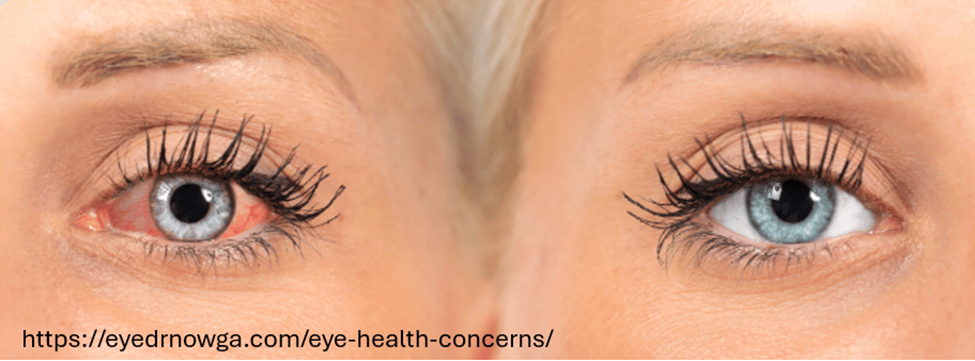
Many times during the day, I let my shoulders and face relax, and with each exhalation I feel the upper eyelids slightly dropping down at the same time as I am relaxing my jaw and mouth while sitting tall. I sense my tongue and throat sinking and feeling an increase in the space between my upper and lower molars. At the same time, I sense my eyes becoming soft and sinking down as my face muscle relax and are pulled down by gravity while sensing a gentle smile. My eyes are not trying to focus on anything. I continue to breathe slowly allowing a pause before inhaling and feel totally safe and at peace. While doing this, I sense moisture at the lower eyelids. After I have inhaled and as I exhale, I slowly open my eyes, and return to my work. My eyes feel slightly moist and as I blink, the eyelids glide smoothly over the corneal surface.
Background
Dry eyes and eye strain are far more common than most people realize. More than 50% of adults in the United States and Europe experience irritated or burning eyes, dryness, eye strain, headaches, tired or heavy eyes, sensitivity to bright light, and general eye discomfort (Wozniak et al., 2025). The prevalence increases with age and is higher among people who smoke, wear contact lenses, or spend excessive time looking at screens—typically more than six hours per day (Uchino et al,, 2013).
Prolonged, intense screen use—often referred to as digital eye strain or computer vision syndrome—occurs when people focus and concentrate, and the muscles involved with near vision contract and do not relax (Chu et al., 2014). During near vision, the ciliary muscles tighten around the lens to allow near focus, and the medial rectus muscles contract to converge the eyes. These muscles stay contracted and only relax when looking into the distance. This ongoing tension and increased sympathetic activation combined with reduced blinking decrease tearing, and contribute to dry eye symptoms because the tears are not able to provide adequate moisture. As a result, the eyes may become irritated, red and inflamed, which increases eye discomfort (Sheppard & Wolffsohn, 2018; Portello et al., 2012; Sheedy et al., 2003).
When someone is vigilant, fearful, or anticipates a threat, sympathetic activation increases (Ranti et al., 2020) and “eye blink are inhibited at precise moments in time so as to minimize the loss of visual information that occurs during a blink. The more important the visual information is to the viewer, the more likely he or she will be to inhibit blinking” (Ranti et al., 2020).
Without being aware, many people are in a chronic state of vigilance and unknowingly are scanning the world for threats to which they must react. Clinically, I often observe this when guiding clients in relaxation. For example, when I give the instruction, “Let me lift your hand,” some clients immediately lift their hand towards me. This nonverbal response suggests that they are constantly vigilant and are monitoring the world around them—always ready to act instead of trusting that they do not have to act and that the world is safe. In most cases, their breathing pattern tends to be shallow and thoracic.
Would it be possible that this ongoing vigilance,which increases sympathetic arousal contributes to the experience of dry eye syndrome? It could be one factor explaining why women have a higher incidence of dry eye disease as well as anxiety than men since the world is often less safe for women, and they breathe more thoracically and less diaphragmatically (abdominally) than men. Shallow chest breathing is also associated with increased in anxiety (Fugl-Meyer, 1974; Mendes et al., 2020; Wilhelm et al., 2001; Banushi eat al., 2023; McLean et al., 2011; Jalnapurkar, et al., 2018).
Note: The risk of dry eye can also be further increased by a wide range of medical conditions and medications, such as diabetes; glaucoma and glaucoma medications; allergies; autoimmune diseases; arthritis; thyroid disease; high cholesterol; acne treatments; antihistamines; antidepressants; and a history of refractive surgery, conjunctival infections, or corneal abrasions. Dry eye also occurs more frequently in women (Mohamed et al., 2024).
What is usually recommended to resolve dry eyes
The primary and effective first-line treatment to reduce the symptoms of dry eye disease is the use of artificial tears (lubricating eye drops) to provide relief (Maity et al., 2025). Another recommendation to reduce eye strain associated with computer use is to implement the 20/20/20 rule developed by the American Optometric Association–take a 20-second break every 20 minutes and look at something 20 feet away (AOA, 2026). Although the 20/20/20 practice can reduce eye strain and momentarily reduce some of the symptoms associated with dry eye disease especially if the eyes are closed, how we orient, breathe and look may contribute to the experience of dry eye discomfort.
Experience how to evoke dry eyes
Sit comfortably, look around, and observe how your eyes feel. Close your eyes. Now imagine there is a threat. Take a very quick gasp through an open mouth by inhaling into your upper chest and keep breathing very shallowly and irregularly. At the same time, open your eyes wide while being vigilant and looking for danger. Simultaneously, tense your body, inclining it slightly backward as if trying to avoid something. Do not blink, as you may miss the potential threat approaching you (adapted from Lemeignan et al., 1990; Bloch, 2017). A sample physiological recording of this pattern is shown in Figure 1.
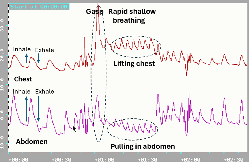
Figure 1. Breathing pattern in response to a threat; a rapid gasp into the chest and pulling the abdomen in to protect while breathing shallowly and rapidly.
Most participants report that almost immediately they feel their eyes getting cooler and after 15 seconds, drier. This facial, breathing, and posture pattern is an exaggeration of the somatic expression of fear that is evoked when we are vigilant and feel unsafe, and it increases sympathetic arousal (Kalawski, 2020).
In most cases, this pattern is automatic, and occurs without awareness, and increases sympathetic activity (Narkiewicz et al., 2006). On the other hand, slow diaphragmatic breathing tends to reduce sympathetic activity (Harada et al., 2014; Lehrer & Gevirtz, 2014). Clinically, the vigilance pattern can often be observed as a person slightly lifts and expands their chest during inhalation and drops and constricts it during exhalation, while breathing shallowly and rapidly without any abdomen expansion or constriction. It is often punctuated with brief breath holding and a reduced blinking rate during concentration.
To investigate these observations more systematically, we compared the practice of gasping while opening the eyes with gentle exhalation while opening the eyes—a pattern that may reduce sympathetic activation and alter the subjective sensation of eye dryness.
Participants: 13 males and 13 females; average age, 39 years
Procedure: While sitting comfortably with their eyes closed, participants were guided through the following two practices:
1.Gasp while opening the eyes
Sit comfortably and look around and observe how your eyes feel. Now close your eyes. Now imagine there is a threat. Take a very quick gasp through an open mouth by inhaling into your upper chest and keep breathing very shallowly and irregularly. At the same time, open your eyes wide while being vigilant and looking for danger. Simultaneously, tense your body, inclining it slightly backward as if trying to avoid something. Do not blink, as you may miss the potential threat approaching you. Repeat three times.
2. Gentle exhalation while opening the eyes
Sit comfortably and look around and observe how your eyes feel. Now close your eyes. Breathe comfortably and inhale by allowing your abdomen to extend and widen while feeling your eyes sinking in their sockets and becoming softer as you gently start exhaling. While gently exhaling, begin to open your eyes very slowly, looking down through your eyelashes without caring what you see, allowing your jaw and face muscles to relax and feeling a slight smile. When you feel the urge to inhale, allow your eyes to close and let your abdomen expand as you inhale slowly. Repeat three times.
After these two practices, the participants filled out a short assessment questionnaire in which they rated how their eyes felt on a scale from -5 (dry), 0 (normal), to 5 (moist/tearing), and rated which eye-opening procedure allowed their eyes to be more relaxed and moist.
Results
92.3% of the participants reported that opening their eyes during exhalation significantly increased eye relaxation and moisture, as shown in Figure 2. A one-way analysis of variance (ANOVA) revealed a significant difference between the inhale condition (M = −0.19, n = 26) and the exhale condition (M = 1.00, n = 25), F(1, 49) = 9.65, p = .003. The experience of eye irritation was correlated (r = 0.64) with the self-rating of experiencing anxiety and fear during the last three months.
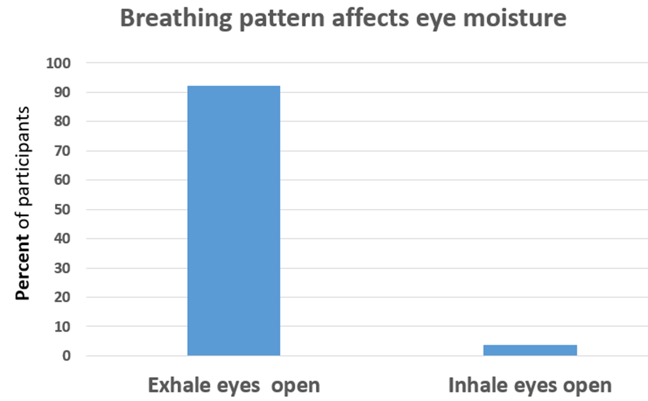
Figure 2. Gently opening the eyes during exhalation increases the experience of moisture and relaxation in the eyes.
Discussion
The results suggest that increased moisture and eye relaxation could be evoked by changing breathing-blinking patterns. Nearly all participants experienced an increase in tearing and eye relaxation; however, long-term benefits most likely occur if the person implements this practice many times during the day. By changing the breathingg-blink pattern and peacefully looking at the world with a smile, one would decrease sympathetic arousal and increase parasympathetic activity. This approach should be taught as the first self-care intervention to reduce eye irritation.
Recommendations to decrease dry eye and improve eye health
- Experience how the two different breathing and eye-opening practices described above affect your eye dryness or moistness.
- When you sense the first onset of minimal eye discomfort or when looking at screens on your computer or cellphone, implement the following practice for about 10 to 15 seconds. Allow your eyelids to close. Be aware of the sensations in your eyes, let your face and jaw relax as if they are being pulled down by gravity while sensing the eyes becoming soft and sinking into their sockets. Breathe diaphragmatically by allowing the abdomen to expand when you inhale. While gently exhaling, slowly open your eyes slightly while looking down with a gentle smile and sensing the moisture beginning to occur in the eyes. Repeat twice.
- During the day, implement the 20/20/20 vision-regeneration practice (every 20 minutes, take a 20-second break and look at something 20 feet away without caring what you see), as shown in Figure 3.
- Read and incorporated the many practices described in the superb book, Vision for Life; Ten Steps to Natural Eye Improvement, by Meir Schneider (2012).
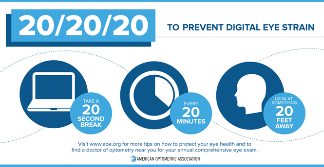
Figure 3. The 20/20/20 rule poster to prevent eye strain (AOA, 2026).
Do these practices many times and be aware of the sensations in your eyes and face. This passive awareness, in conjunction with slower breathing, tends to reduce sympathetic arousal and increase parasympathetic activity, which facilitates increased tearing. As one participant reported when she practiced this:
What a surprise it was when I closed my eyes and breathed slowly and diaphragmatically, and then, as I began to exhale, I very slowly began to open my eyes while looking down and through my eyelashes, while feeling my eyes sink into their sockets. Tearing occurred spontaneously, and my eyes felt lubricated. What a relief. It provided hope that I could help myself instead of only depending on lubricating eye drops whenever my eyes felt dry and irritated.
Two books to maintain and improve vision and reduce techstress
Vision for Life: Ten Steps to Natural Eye Improvement by Meir Schneider (2016) offers many strategies you can immediately incorporate into your daily life to improve and restore your vision. It provides guidelines on how to reverse developing vision issues before they cause damage and how to remedy existing problems, including near- and far-sightedness, lazy eye, as well as more serious conditions such as cataracts, glaucoma, optic neuritis, detached retinas and retinal tears, macular degeneration, and retinitis pigmentosa.
TechStress: How Technology is Hijacking our Lives, Strategies for Coping, and Pragmatic Ergonomics by Erik Peper, Richard Harvey, and Nancy Faass (2020) offers practical tools to avoid the evolutionary traps that trip us up and address the problems associated with technological overuse. It includse effective strategies and practices that individuals can use to optimize their workspace, reduce physical strain, correct posture, and improve vision. It provides fresh insights on reducing stress and enhancing health.
Listen to the expanded podcast based on this blog produced with Google Notebook LM.
Additional blogs that offer strategies to improve vision
References
AOA. (2026). 20/20/20/ to prevent digital eyes strain. American Optometric Association. https://www.aoa.org/AOA/Images/Patients/Eye%20Conditions/20-20-20-rule.pdf
Banushi, B., Brendle, M., Ragnhildstveit, A., Murphy, T., Moore, C., Egberts, J., & Robison, R. (2023). Breathwork interventions for adults with clinically diagnosed anxiety disorders: A scoping review. Brain Sciences, 13(2), 256. https://doi.org/10.3390/brainsci13020256
Bloch, S. (2017). Alba Emoting: A scientific method for emotional induction. Scotts Valley, CA: CreateSpace Independent Publishing Platform. https://www.amazon.com/Alba-Emoting-Scientific-Emotional-Induction/dp/154254884
Chu, C.A., Rosenfield, M., Portello, J.K. (2014). Blink patterns: reading from a computer screen versus hard copy. Optom Vis Sci., 91(3),297-302. https://doi.org/10.1097/OPX.0000000000000157
Craig, J. P., Nichols, K. K., Akpek, E. K., et al. (2017). TFOS DEWS II definition and classification report. The Ocular Surface, 15(3), 276–283. https://doi.org/10.1016/j.jtos.2017.05.008
Dartt, D. A. (2009). Neural regulation of lacrimal gland secretory processes: Relevance in dry eye diseases. Progress in Retinal and Eye Research, 28(3), 155–177. https://doi.org/10.1016/j.preteyeres.2009.04.003
Fugi-Meyer, A.R. (1974). Relative respiratory contribution of the rib cage and the abdomen in males and females with special regard to posture. Respiration, 31(3), 240–251. https://doi.org/10.1159/000193113
Harada, D., Asanoi, H., Takagawa, J., Ishise, H., Ueno, H., Oda, Y., Goso, Y., Joho, S., & Inoue, H. (2014). Slow and deep respiration suppresses steady-state sympathetic nerve activity in patients with chronic heart failure: from modeling to clinical application. American Journal of Physiology-Heart and Circulatory Physiology, 307(8), H1159–H1168. https://doi.org/10.1152/ajpheart.00109.2014
Jalnapurkar, I., Allen, M., & Pigott, T. (2018). Sex differences in anxiety disorders: A review. Journal of Psychiatry, Depression & Anxiety, 4, 011. https://doi.org/10.24966/PDA-0150/100011
Kalawski, J.P. (2020) The Alba Method and the Science of Emotions. Integr. Psych. Behav. 54, 903–919. https://doi.org/10.1007/s12124-020-09525-4
Lehrer, P. M., & Gevirtz, R. (2014). Heart rate variability biofeedback: How and why does it work? Frontiers in Psychology, 5, Article 756. https://www.ncbi.nlm.nih.gov/pmc/articles/PMC4104929/
Lemeignan, M., Guitart, L., & Bloch, S. (1990). Autonomic differentiation of emotional effector pattern of 6 basic emotions. Proceedings of the Fifth International Congress of Psychophysiology, Budapest, July 9–14, 199
Maity, M., Allay, M. B., Ali, M. H., Basu, S., & Singh, S. (2025). Effect of different artificial tears on tear film parameters in dry eye disease. Optometry and Vision Science, 102(1), 37–43. https://doi.org/10.1097/OPX.0000000000002206
McLean, C. P., Asnaani, A., Litz, B. T., & Hofmann, S. G. (2011). Gender differences in anxiety disorders: Prevalence, course of illness, comorbidity and burden of illness. Journal of Psychiatric Research, 45(8), 1027–1035. https://doi.org/10.1016/j.jpsychires.2011.03.006
Mendes, L. P. S., Vieira, D. S. R., Gabriel, L. S., Ribeiro-Samora, G. A., Dornelas de Andrade, A., Brandão, D. C., Goes, M. C., Fregonezi, G. A. F., Britto, R. R., & Parreira, V. F. (2020). Influence of posture, sex, and age on breathing pattern and chest wall motion in healthy subjects. Brazilian Journal of Physical Therapy, 24(3), 240–248. https://doi.org/10.1016/j.bjpt.2019.02.007
Mohamed, Z., Alrasheed, S., Abdu, M., & Allinjawi, K. (2024). Dry eye disease prevalence and associated risk factors among the Middle East population: A systematic review and meta-analysis. Cureus, 16(9), e70522. https://doi.org/10.7759/cureus.70522
Narkiewicz, K., van de Borne, P., Montano, N., Hering, D., Kara, T., & Somers, V. K. (2006). Sympathetic neural outflow and chemoreflex sensitivity are related to spontaneous breathing rate in normal men. Hypertension, 47(1), 51–55. https://doi.org/10.1161/01.HYP.0000197613.47649.0
Peper, E., Harvey, R., & Faass, N. (2020). TechStress: How Technology is Hijacking our Lives, Strategies for Coping, and Pragmatic Ergonomics, North Atlantic Books. https://www.amazon.com/Beyond-Ergonomics-Prevent-Fatigue-Burnout/dp/158394768X
Portello, J.K., Rosenfield, M., Bababekova, Y., et al. (2012). Computer-related visual symptoms in office workers. Ophthalmic and Physiological Optics, 32, 375–82. https://doi.org/10.1111/j.1475-1313.2012.00925.x
Ranti, C., Jones, W., Klin, A. et al. Blink Rate Patterns Provide a Reliable Measure of Individual Engagement with Scene Content. Sci Rep 10, 8267 (2020). https://doi.org/10.1038/s41598-020-64999-x
Schneider, M. (2016). Vision for Life: Ten Steps to Natural Eye Improvement. North Atlantic Books. https://www.amazon.com/Vision-Life-Revised-Eyesight-Improvement/dp/1623170087
Sheedy, J.E., Hayes, J.N., & Engle, J.(2003). Is all asthenopia the same? Optom Vis Sci, 80, 732–9. https://doi.org/10.1097/00006324-200311000-00008
Sheppard, A. L., & Wolffsohn, J. S. (2018). Digital eye strain: prevalence, measurement and amelioration. BMJ open Ophthalmology, 3(1), e000146. https://doi.org/10.1136/bmjophth-2018-000146
Stern, M. E., Gao, J., Siemasko, K. F., Beuerman, R. W., & Pflugfelder, S. C. (2004). The role of the lacrimal functional unit in the pathophysiology of dry eye. Experimental Eye Research, 78(3), 409–416. https://doi.org/10.1016/j.exer.2003.09.003
Swamynathan, S. K., & Wells, A. (2020). Conjunctival goblet cells: Ocular surface functions, disorders that affect them, and the potential for their regeneration. The Ocular Surface, 18(1), 19–26. https://doi.org/10.1016/j.jtos.2019.11.005
Uchino, M., Yokoi, N., Uchino, Y., Dogru, M., Kawashima, M., Komuro, A., Sonomura, Y., Kato, H., Kinoshita, S., Schaumberg, D.A., & Tsubota, K. (2013). Prevalence of Dry Eye Disease and its Risk Factors in Visual Display Terminal Users: The Osaka Study. American Journal of Ophthalmology, 156(4), 759-766.e1, https://doi.org/10.1016/j.ajo.2013.05.040
The epidemiology of dry eye disease: report of the Epidemiology Subcommittee of the International Dry Eye WorkShop (2007). Ocul Surf. , 5(2), 93-107. https://doi.org/10.1016/s1542-0124(12)70082-4
Wilhelm, F. H., Gevirtz, R., & Roth, W. T. (2001). Respiratory dysregulation in anxiety, functional cardiac, and pain disorders: Assessment, phenomenology, and treatment. Behavior Modification, 25(4), 513–545. https://doi.org/10.1177/0145445501254003
Wozniak, P., et al. (2025, September 12–16). Dry eye symptoms, severity, treatment and unmet needs: An analysis of the United States of America and a multinational snapshot (NESTS Study) [Poster presentation]. 43rd Congress of the European Society of Cataract and Refractive Surgeons (ESCRS), Copenhagen, Denmark. https://www.sciencedaily.com/releases/2025/09/250914205829.htm
Ziemssen, F., & Ruprecht, K. W. (2005).Autonomic dysfunction in dry eye syndrome. Ophthalmologe, 102(8), 744–749. https://doi.org/10.1007/s00347-005-1169-1
Corporations: The new(old) disease vectors as they choose profits over health
Posted: February 24, 2026 Filed under: Breathing/respiration, cancer, health | Tags: cancer, environment, environmental toxins, food, health, herbicides, investigative reporting, nutrition, pollution, public health, smoking, wellness 1 Comment
“We can invest in preventing illness now by reducing our exposure to environmental toxins — or we can pay a far higher price later trying to treat the resulting chronic and often debilitating diseases.”
Ever since the 1962 publication of Rachel Carson’s groundbreaking book, Silent Spring, which documented the harm environmental pollution caused, government has, often reluctantly, set limits intended to protect Americans from exposure to harmful chemicals in our food, air and water (Carson, 1964). These regulations did not emerge easily. As the governmental regulations were being proposed and implemented, they were consistently challenged by the very large corporations that manufactured and profited from these chemicals.
History reminds us how slowly public health protections can unfold. Consider how long it took for smoking to be prohibited in public spaces even though the harmful effects had been documented since the 1950s (Doll and Hill, 1954; Doll & Hill, 1964; Wynder & Graham,1985). For decades, the science was clear, yet policy and governmental actions were delayed. Only in the early 2000s did many states began banning smoking in workplaces, restaurants, and bars. The shift in public policy saved many lives and the reduction in smoking has been the major reason for the decrease in cancer mortality over the last twenty-five years.
We are going backwards
The Trump administration has rescinded the 2009 U.S. Environmental Protection Agency endangerment finding on greenhouse gases, loosening vehicle emission standards, and weakening pollution controls on power plants and oil and gas operations (Tabuchi, 2026). The health consequences may not appear immediately; however, they are predictable. The increased exposure today will again contribute to increased rates of cancer, respiratory illness, cardiovascular disease, and developmental disorders tomorrow.
To understand how the government regulations have been revised so that once again Americans will be more exposed to toxins in their food, air, and water, read the superb investigative report published by U.S. Right to Know whose mission is to pursuing truth and transparency for public health.
Their most recent report, Tracing Bayer’s ties to power in Trump’s Washington, describes in detail the hidden social connections, lobbying and political donations that lead “The White House to invokes the Defense Production Act to guarantee supplies of elemental phosphorus and glyphosate-based herbicides. Regulators reapprove dicamba, a Bayer herbicide twice blocked by federal courts, and clear the way for new pesticides containing toxic, persistent PFAS “forever” chemicals (Malkan, 2026).“
When regulatory safeguards weaken, corporations can once again function as disease vectors-not through infection, but through environmental exposure. By loosening the pollution standards, federal policy will negatively affect the health of both present and future generations.
I encourage you to explore many superb investigative reports and practical suggestions how to avoid these toxins exposure that are available on U.S. Right to Know website:
One exposure. Twenty generations later, the damage is still unfolding
Glyphosate: Cancer, liver disease, endocrine disruption and other health concerns
Big Food ‘transparency’ campaign seeks to block tough new food safety laws
Ultra-processed foods damage health in ways that calories don’t explain, new study says
Listen to the expanded podcast based upon this blog and created by Google Notebook LM.
See also the following blogs
References
Carson, R. (1962). Silent spring. Houghton Mifflin. https://www.amazon.com/Silent-Spring-Rachel-Carson/dp/B002E8JF6G/
Doll, R., & Hill, A. B. (1954). The mortality of doctors in relation to their smoking habits: A preliminary report. British Medical Journal, 1(4877), 1451–1455. https://doi.org/10.1136/bmj.1.4877.1451
Doll, R., & Hill, A. B. (1964). Mortality in relation to smoking: Ten years’ observations of British doctors. British Medical Journal, 1(5396), 1460–1467. https://doi.org/10.1136/bmj.1.5396.1460
Malkan, S. (2026). Tracing Bayer’s ties to power in Trump’s Washington. U.S. Right to Know. Accessed February 24, 2026. https://usrtk.org/pesticides/tracing-bayers-ties-to-power-in-trumps-washington/
Tabuchi, H. (2026). Historic Climate Rollback Makes U.S. a Global Outlier on Tailpipe Rules. The New York Times, February 16, 2026. Accessed February 24, 2026 https://www.nytimes.com/2026/02/16/climate/endangerment-finding-auto-emissions-regulations.html
Wynder, E.L., & Graham, A. (1985). JAMA, 253 (20), 2986-2994. https://doi.org/10.1001/jama.1985.03350440064033